Review
. 2021 Jul 1;32(14):1267-1272.
doi: 10.1091/mbc.E20-10-0681.

Roles of leader and follower cells in collective cell migration

Affiliations
Review

Roles of leader and follower cells in collective cell migration

Lei Qin et al. Mol Biol Cell. .

Abstract

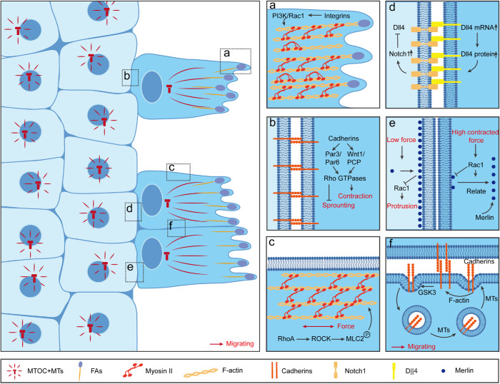
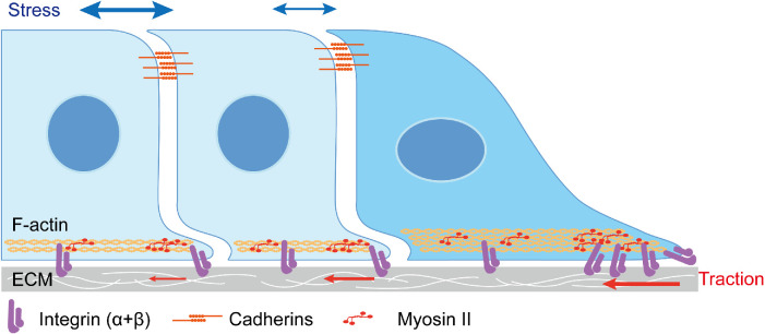
Collective cell migration is a widely observed phenomenon during animal development, tissue repair, and cancer metastasis. Considering its broad involvement in biological processes, it is essential to understand the basics behind the collective movement. Based on the topology of migrating populations, tissue-scale kinetics, called the "leader-follower" model, has been proposed for persistent directional collective movement. Extensive in vivo and in vitro studies reveal the characteristics of leader cells, as well as the special mechanisms leader cells employ for maintaining their positions in collective migration. However, follower cells have attracted increasing attention recently due to their important contributions to collective movement. In this Perspective, the current understanding of the molecular mechanisms behind the "leader-follower" model is reviewed with a special focus on the force transmission and diverse roles of leaders and followers during collective cell movement.


Figures

FIGURE 1:
FIGURE 1:
Differentiation and maintenance of leader cells in collective cell migration. Top view of 2D collective sheet migration. Leader cells are front cells in dark blue with polarized centrosome–nucleus axis orientation and distinct “finger-like” protrusion generated from focal adhesions. Follower cells are the major cell population in light blue located in the cell reservoir with random centrosome–nucleus axis orientation and low migratory speed. (A) At the tip of leader cells, large lamellipodia protrusions extend from the cell body and form a “finger-like” morphology. At these sites, strong integrin-based FA connections with ECM activate a downstream PI3K-Rac signaling pathway, which further enhances actomyosin bundle formation and traction force generation. (B) At the rear of the leader cells, transmembrane protein cadherins mediate cell–cell connections with follower cells. At these sites, cadherin-mediated CIL leads to Rho-kinase–dependent myosin light-chain 2 phosphorylation, or Par3/Par6 recruitment at the junctional sites, which result in sprouting inhibition. (C) At the side of leader cells, phosphorylated myosin light chain and F-actin are highly accumulated as thick bundles, which prohibit new protrusion generation from follower cells. (d) Notch1-Dll4 lateral inhibition is reported at the interphase between leader and follower cells, with high Dll4 detected in leader cells, whereas high Notch1 is detected in follower cells. Low cellular stress in leader cells enhances Dll4 mRNA and protein expression, which further determinates the initiation of leader cells and migrating tips. This high Dll4 expression in leader cells enhances the Notch1 expression in follower cells, which in turn inhibits Dll4 expression in these cells. Moreover, high cellular stress also suppresses Dll4 expression in follower cells. (e) Merlin-Rac lateral inhibition is reported at the interphase between leader and follower cells. High contractile forces result in cytoplasmic Merlin through Rac-dependent translocation of Merlin in leader cells, whereas low contractile forces lead to boundary Merlin in follower cells which further inhibit Rac activity and Rac-mediated protrusions in follower cells. (F) At the interphase of lateral membranes between two migrating leader cells, a continuous treadmilling of cadherins is achieved through GSK3-dependent endocytosis processes.
FIGURE 2:
FIGURE 2:
Mechanical force distribution in the “leader–follower” model. Lateral view of 2D collective sheet migration. Leader cells in dark blue exhibit asymmetry exposure of adhesions, which includes integrin-based FA at migratory front and cadherin-mediated AJ at the rear with follower cells. Whereas, follower cells in light blue experience symmetry cadherin-mediated AJ with neighboring cells at their apical surface and small FA formation at their basal surface. From the mechanical force perspective, the collective migrating population participates in a global “tug-of-war.” The highly accumulated integrins at the migrating tip enable leader cells with large traction forces generated with ECM, and small tractions are recorded in follower cells with their “cryptic” protrusions. The balanced forces to traction are the cellular stress, which is mainly transmitted by the cellular cytoskeleton, and the cell–cell junctions. The stress is built up across the entire migrating tissue, and increases steadily as the distance from the leading edge increases.

References

    1. Abraham S, Yeo M, Montero-Balaguer M, Paterson H, Dejana E, Marshall CJ, Mavria G (2009). VE-cadherin-mediated cell-cell interaction suppresses sprouting via signaling to MLC2 phosphorylation. Curr Biol 19, 668–674. - PubMed
    1. Alert R, Trepat X (2019). Physical models of collective cell migration. Ann Rev Condens Matter Phys 11, 77–101.
    1. Aman A, Piotrowski T (2010). Cell migration during morphogenesis. Dev Biol 341, 20–33. - PubMed
    1. Aoki K, Kondo Y, Naoki H, Hiratsuka T, Itoh RE, Matsuda M (2017). Propagating wave of ERK activation orients collective cell migration. Dev Cell 43, 305–317.e5. - PubMed
    1. Barriga EH, Franze K, Charras G, Mayor R (2018). Tissue stiffening coordinates morphogenesis by triggering collective cell migration in vivo. Nature 554, 523–527. - PMC - PubMed

Publication types

LinkOut - more resources