. 2022 Jan;26(1):609-622.
doi: 10.1007/s00784-021-04039-8. Epub 2021 Jun 29.

Cyclic tensile strain affects the response of human periodontal ligament stromal cells to tumor necrosis factor-α

Affiliations

Cyclic tensile strain affects the response of human periodontal ligament stromal cells to tumor necrosis factor-α

Zhongqi Zhao et al. Clin Oral Investig. 2022 Jan.

Abstract

Objectives: Orthodontic treatment in adult patients predisposed to mild or severe periodontal disease is challenging for orthodontists. Orthodontic malpractice or hyper-occlusal forces may aggravate periodontitis-induced destruction of periodontal tissues, but the specific mechanism remains unknown. In the present study, the combined effect of mechanical stress and tumor necrosis factor (TNF)-α on the inflammatory response in human periodontal ligament stromal cells (hPDLSCs) was investigated.

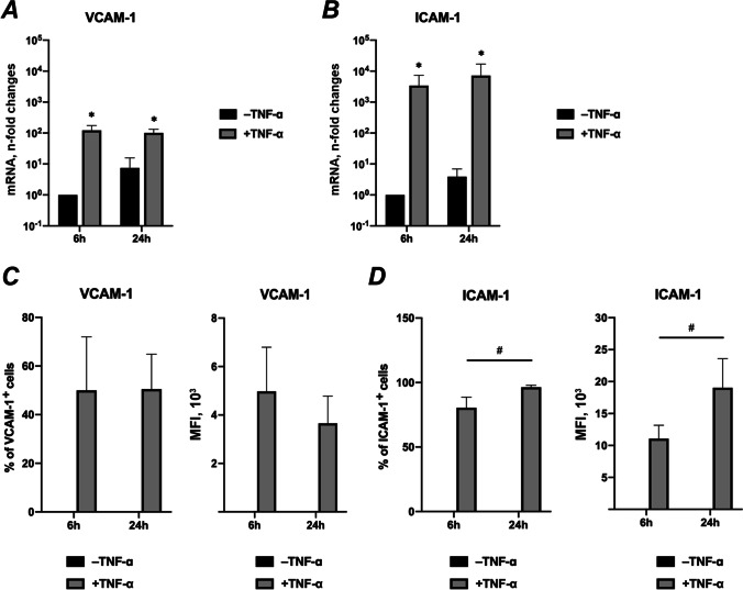
Materials and methods: hPDLSCs from 5 healthy donors were treated with TNF-α and/or subjected to cyclic tensile strain (CTS) of 6% or 12% elongation with 0.1 Hz for 6- and 24 h. The gene expression of interleukin (IL)-6, IL-8 and cell adhesion molecules VCAM and ICAM was analyzed by qPCR. The protein levels of IL-6 and IL-8 in conditioned media was measured by ELISA. The surface expression of VCAM-1 and ICAM-1 was quantified by immunostaining followed by flow cytometry analysis.

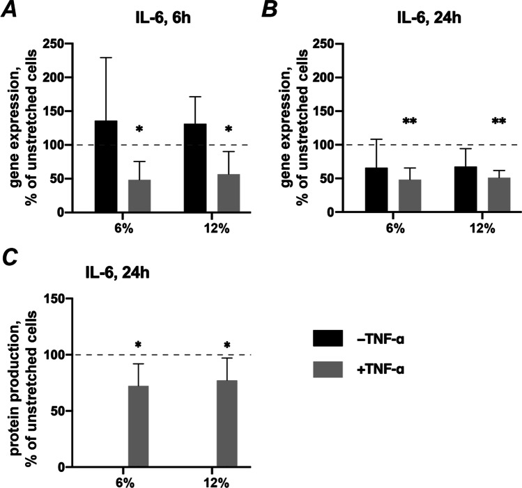
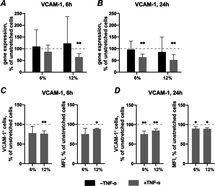
Results: TNF-α-induced IL-6 gene and protein expression was inhibited by CTS, whereas TNF-α-induced IL-8 expression was decreased at mRNA expression level but enhanced at the protein level in a magnitude-dependent manner. CTS downregulated the gene expression of VCAM-1 and ICAM-1 under TNF-α stimulation, but the downregulation of the surface expression analyzed by flow cytometry was observed chiefly for VCAM-1.

Conclusions: Our findings show that mechanical force differentially regulates TNF-α-induced expression of inflammatory mediators and adhesion molecules at the early stage of force application. The effect of cyclic tensile strain is complex and could be either anti-inflammatory or pro-inflammatory depending on the type of pro-inflammatory mediators and force magnitude.

Clinical relevance: Orthodontic forces regulate the inflammatory mediators of periodontitis. The underlying mechanism may have significant implications for future strategies of combined periodontal and orthodontic treatment.

Keywords: Human periodontal ligament stromal cells; Inflammatory cytokine; Mechanical loading; Orthodontic force; Periodontitis.


Conflict of interest statement

The authors declare no competing interests.

Figures

Fig. 1
Fig. 1
Schematic of mechanical force loading. (a) BioFlex® Cell Seeders were applied before seeding to confine the cell suspension in the central area of a well in the culture plates. (b) The culture plates were transferred onto the loading post after seeding hPDLSCs at a density of 1 × 105 cells/well in the central region of the flexible membrane using BioFlex® Cell Seeders. (c) Cells were stretched by a cyclic tensile strain (equibiaxial, 6 or 12% elongation, sinusoidal curve, 0.1 Hz, 6 or 24 h) when the vacuum was applied
Fig. 2
Fig. 2
Effects of TNF-α on gene and protein expression of IL-6 and IL-8 on unstretched hPDLSCs. Primary hPDLSCs were treated with 10 ng/ml TNF-α without mechanical stretching. After 6- or 24 h of incubation, gene expression levels of IL-6 (a), IL-8 (b) were measured using RT-qPCR. The Y-axes show the n-fold changes in mRNA expression compared to untreated control after 6 h (n-fold expression = 1). The corresponding protein levels of IL-6 (c) and IL-8 (d) in conditioned media were quantified by ELISA. The groups in the absence or presence of TNF-α are indicated as -TNF-α or + TNF-α, respectively. The data are presented as the mean ± standard deviation. *p < 0.05 compared to corresponding control. #p < 0.01 compared between groups as indicated
Fig. 3
Fig. 3
Effects of CTS on IL-6 expression in the absence / presence of TNF-α in hPDLSCs. Primary hPDLSCs were subjected to either 6% or 12% cyclic stretching, in the absence or presence of 10 ng/ml TNF-α for 6 or 24 h. IL-6 gene expression levels were measured with RT-qPCR after 6 (a) or 24 (b) hours. TNF-α-induced IL-6 protein levels after 24 h (c) were quantified by ELISA. Y-axes show the effect of CTS on IL-6 expression as % of the values observed in unstretched cells with or without TNF-α. For RT-qPCR (a, b), n-fold gene expression was calculated first using the 2−ΔΔCt method and then the data were normalized to those observed in unstretched cells (100%, dashed line). For ELISA (c), the values were calculated as % of the values measured in unstretched cells (100%, dashed line). The data are presented as the mean ± standard deviation. * and ** — significantly different compared to unstretched control with p < 0.05 and p < 0.01, respectively
Fig. 4
Fig. 4
Effects of CTS on IL-8 expression in the absence / presence of TNF-α in hPDLSCs. Primary hPDLSCs were subjected to either 6% or 12% cyclic stretching, in the absence or presence of 10 ng/ml TNF-α. IL-8 gene expression levels were measured with RT-qPCR after 6 (a) or 24 (b) hours. TNF-α-induced IL-8 protein levels after 6 (c) and 24 (d) hours were quantified by ELISA. Y-axes show the effect of CTS on IL-8 expression as % of the values observed in unstretched cells with or without TNF-α. For RT-qPCR (a, b), n-fold gene expression was calculated first using the 2−ΔΔCt method and then the data were normalized to those observed in unstretched cells (dashed line). For ELISA (c, d), the values were calculated as % of the values measured in unstretched cells (100%, dashed line). The data are presented as the mean ± standard deviation. * and ** — significantly different compared to unstretched control with p < 0.05 and p < 0.01, respectively
Fig. 5
Fig. 5
Effects of TNF-α on gene and protein expression of VCAM-1 and ICAM-1 in unstretched hPDLSCs. Primary hPDLSCs were treated with 10 ng/ml TNF-α without mechanical stretching. The groups in the absence or presence of TNF-α are indicated as -TNF-α or + TNF-α, respectively. After 6- or 24 h of incubation, gene expression levels of VCAM-1 (a) and ICAM-1 (b) were measured using RT-qPCR. The Y-axes show the n-fold changes in mRNA expression compared to untreated control after 6 h (n-fold expression = 1). The corresponding protein levels of VCAM-1 (c) and ICAM-1 (d) were quantified by flow cytometry. The Y-axes show the percentage of positive cells and the corresponding mean fluorescence intensity (MFI) of the positive cell population, respectively. No positively stained VCAM-1 and ICAM-1 cells were observed in the absence of TNF-α. The data are presented as the mean ± standard deviation. *p < 0.05 compared to corresponding control. #p < 0.01 compared between groups as indicated
Fig. 6
Fig. 6
Effects of CTS on VCAM-1 expression in the absence/presence of TNF-α in hPDLSCs. Primary hPDLSCs were subjected to either 6% or 12% cyclic stretching, in the absence or presence of 10 ng/ml TNF-α. VCAM-1 gene expression levels were measured with RT-qPCR using the 2−ΔΔCt method after 6 (a) or 24 (b) hours. TNF-α-induced VCAM-1 protein levels after 6 (c) and 24 (d) hours were quantified by flow cytometry. Y-axes show the effect of CTS on VCAM-1 expression as % of the values observed in unstretched cells with or without TNF-α. For RT-qPCR (a, b), n-fold gene expression was calculated first using the 2−ΔΔCt method and then the data were normalized to those observed in unstretched cells (dashed line). For protein expression (c, d), Y-axes show the percentage of positive cells and the mean fluorescence intensity (MFI) of the positive cell population, in % of the values measured in unstretched cells. The data are presented as the mean ± standard deviation. * and ** — significantly different compared to unstretched control with p < 0.05 and p < 0.01, respectively
Fig. 7
Fig. 7
Effects of CTS on ICAM-1 expression in the absence / presence of TNF-α in hPDLSCs. Primary hPDLSCs were subjected to either 6% or 12% cyclic stretching, in the absence or presence of 10 ng/ml TNF-α. Unstretched hPDLSCs served as control. ICAM-1 gene expression levels were measured with RT-qPCR after 6 (a) or 24 (b) hours. TNF-α-induced ICAM-1 protein levels after 6 (c) and 24 (d) hours were quantified by flow cytometry. Y-axes show the effect of CTS on ICAM-1 expression as % of the values observed in unstretched cells with or without TNF-α. For RT-qPCR (a, b), n-fold gene expression was calculated first using the 2−ΔΔCt method and then the data were normalized to those observed in unstretched cells (dashed line). For protein expression (c, d) the Y-axes show the percentage of positive cells and the mean fluorescence intensity (MFI) of the positive cell population, in % of the values measured in unstretched cells. The data are presented as the mean ± standard deviation. * and ** — significantly different compared to unstretched control with p < 0.05 and p < 0.01, respectively

References

    1. Proffit WR (2018) Contemporary orthodontics. 6th edition. edn. Elsevier, Philadelphia, IL
    1. McCulloch CA, Lekic P, McKee MD. Role of physical forces in regulating the form and function of the periodontal ligament. Periodontol. 2000;24:56–72. doi: 10.1034/j.1600-0757.2000.2240104.x. - DOI - PubMed
    1. Jin Y, Ding L, Ding Z, Fu Y, Song Y, Jing Y, Li Q, Zhang J, Ni Y, Hu Q. Tensile force-induced PDGF-BB/PDGFRbeta signals in periodontal ligament fibroblasts activate JAK2/STAT3 for orthodontic tooth movement. Sci Rep. 2020;10(1):11269. doi: 10.1038/s41598-020-68068-1. - DOI - PMC - PubMed
    1. Behm C, Nemec M, Blufstein A, Schubert M, Rausch-Fan X, Andrukhov O, Jonke E. Interleukin-1beta induced matrix metalloproteinase expression in human periodontal ligament-derived mesenchymal stromal cells under in vitro simulated static orthodontic forces. Int J Mol Sci. 2021;22:3. doi: 10.3390/ijms22031027. - DOI - PMC - PubMed
    1. Ivanovski S, Gronthos S, Shi S, Bartold PM. Stem cells in the periodontal ligament. Oral Dis. 2006;12(4):358–363. doi: 10.1111/j.1601-0825.2006.01253.x. - DOI - PubMed

Substances