Pathogenic MAST3 Variants in the STK Domain Are Associated with Epilepsy
- PMID: 34185323
- PMCID: PMC8324566
- DOI: 10.1002/ana.26147
Pathogenic MAST3 Variants in the STK Domain Are Associated with Epilepsy
Abstract
Objective: The MAST family of microtubule-associated serine-threonine kinases (STKs) have distinct expression patterns in the developing and mature human and mouse brain. To date, only MAST1 has been conclusively associated with neurological disease, with de novo variants in individuals with a neurodevelopmental disorder, including a mega corpus callosum.
Methods: Using exome sequencing, we identify MAST3 missense variants in individuals with epilepsy. We also assess the effect of these variants on the ability of MAST3 to phosphorylate the target gene product ARPP-16 in HEK293T cells.
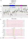
Results: We identify de novo missense variants in the STK domain in 11 individuals, including 2 recurrent variants p.G510S (n = 5) and p.G515S (n = 3). All 11 individuals had developmental and epileptic encephalopathy, with 8 having normal development prior to seizure onset at <2 years of age. All patients developed multiple seizure types, 9 of 11 patients had seizures triggered by fever and 9 of 11 patients had drug-resistant seizures. In vitro analysis of HEK293T cells transfected with MAST3 cDNA carrying a subset of these patient-specific missense variants demonstrated variable but generally lower expression, with concomitant increased phosphorylation of the MAST3 target, ARPP-16, compared to wild-type. These findings suggest the patient-specific variants may confer MAST3 gain-of-function. Moreover, single-nuclei RNA sequencing and immunohistochemistry shows that MAST3 expression is restricted to excitatory neurons in the cortex late in prenatal development and postnatally.
Interpretation: In summary, we describe MAST3 as a novel epilepsy-associated gene with a potential gain-of-function pathogenic mechanism that may be primarily restricted to excitatory neurons in the cortex. ANN NEUROL 2021;90:274-284.
© 2021 American Neurological Association.
Conflict of interest statement
Potential conflicts of interest
The authors report no competing interests.
Figures


Similar articles
-
The Role of Microtubule Associated Serine/Threonine Kinase 3 Variants in Neurodevelopmental Diseases: Genotype-Phenotype Association.Front Mol Neurosci. 2022 Jan 12;14:775479. doi: 10.3389/fnmol.2021.775479. eCollection 2021. Front Mol Neurosci. 2022. PMID: 35095415 Free PMC article.
-
ARPP-16 Is a Striatal-Enriched Inhibitor of Protein Phosphatase 2A Regulated by Microtubule-Associated Serine/Threonine Kinase 3 (Mast 3 Kinase).J Neurosci. 2017 Mar 8;37(10):2709-2722. doi: 10.1523/JNEUROSCI.4559-15.2017. Epub 2017 Feb 6. J Neurosci. 2017. PMID: 28167675 Free PMC article.
-
Developmental and epilepsy spectrum of KCNB1 encephalopathy with long-term outcome.Epilepsia. 2020 Nov;61(11):2461-2473. doi: 10.1111/epi.16679. Epub 2020 Sep 21. Epilepsia. 2020. PMID: 32954514
-
Neonatal developmental and epileptic encephalopathy due to autosomal recessive variants in SLC13A5 gene.Epilepsia. 2020 Nov;61(11):2474-2485. doi: 10.1111/epi.16699. Epub 2020 Oct 16. Epilepsia. 2020. PMID: 33063863 Review.
-
Trio exome sequencing identified a novel de novo WASF1 missense variant leading to recurrent site substitution in a Chinese patient with developmental delay, microcephaly, and early-onset seizures: A mutational hotspot p.Trp161 and literature review.Clin Chim Acta. 2021 Dec;523:10-18. doi: 10.1016/j.cca.2021.08.030. Epub 2021 Aug 31. Clin Chim Acta. 2021. PMID: 34478686 Review.
Cited by
-
Microtubule-Associated Serine/Threonine (MAST) Kinases in Development and Disease.Int J Mol Sci. 2023 Jul 25;24(15):11913. doi: 10.3390/ijms241511913. Int J Mol Sci. 2023. PMID: 37569286 Free PMC article. Review.
-
Genetic and clinical insights into MAST4-related neurodevelopmental disorders.Front Pediatr. 2025 Jun 27;13:1603050. doi: 10.3389/fped.2025.1603050. eCollection 2025. Front Pediatr. 2025. PMID: 40656202 Free PMC article.
-
Microtubule associated serine/threonine kinase-3 inhibits the malignant phenotype of breast cancer by promoting phosphorylation-mediated ubiquitination degradation of yes-associated protein.Breast Cancer Res. 2025 May 1;27(1):63. doi: 10.1186/s13058-025-02028-3. Breast Cancer Res. 2025. PMID: 40312366 Free PMC article.
-
The Role of Microtubule Associated Serine/Threonine Kinase 3 Variants in Neurodevelopmental Diseases: Genotype-Phenotype Association.Front Mol Neurosci. 2022 Jan 12;14:775479. doi: 10.3389/fnmol.2021.775479. eCollection 2021. Front Mol Neurosci. 2022. PMID: 35095415 Free PMC article.
-
MAST Kinases' Function and Regulation: Insights from Structural Modeling and Disease Mutations.Biomedicines. 2025 Apr 9;13(4):925. doi: 10.3390/biomedicines13040925. Biomedicines. 2025. PMID: 40299535 Free PMC article.
References
-
- McTague A, Howell KB, Cross JH, Kurian MA, Scheffer IE. The genetic landscape of the epileptic encephalopathies of infancy and childhood. Lancet Neurol. 2015. March;15(3):304–16. - PubMed
-
- Scheffer IE. A new classification and class 1 evidence transform clinical practice in epilepsy. Lancet Neurol. 2017. January;17(1):7–8. - PubMed
-
- Garland P, Quraishe S, French P, O’Connor V. Expression of the MAST family of serine/threonine kinases. Brain Research. 2008;1195:12–9. - PubMed
Publication types
MeSH terms
Substances
Grants and funding
- U01 HG007690/HG/NHGRI NIH HHS/United States
- NIH Common Fund
- Office of the NIH Director
- NIH
- R00 NS089858/NS/NINDS NIH HHS/United States
- Japan Agency for Medical Research and Development
- National Health and Medical Research Council
- NH/NIH HHS/United States
- U01 HG007530/HG/NHGRI NIH HHS/United States
- Office of Strategic Coordination
- Yale University
- P50 HD103524/HD/NICHD NIH HHS/United States
- UL1 TR001422/TR/NCATS NIH HHS/United States
- March of Dimes
LinkOut - more resources
Full Text Sources
Medical
Molecular Biology Databases

