. 2021 Aug 9;6(15):e150107.
doi: 10.1172/jci.insight.150107.

IL-13 is a driver of COVID-19 severity

Affiliations

IL-13 is a driver of COVID-19 severity

Alexandra N Donlan et al. JCI Insight. .

Abstract

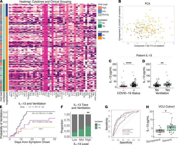
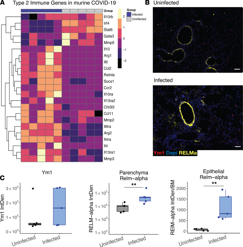
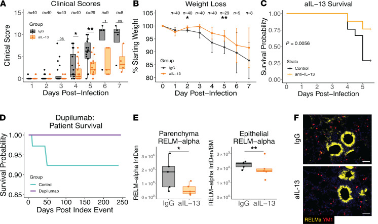
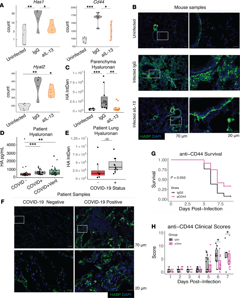
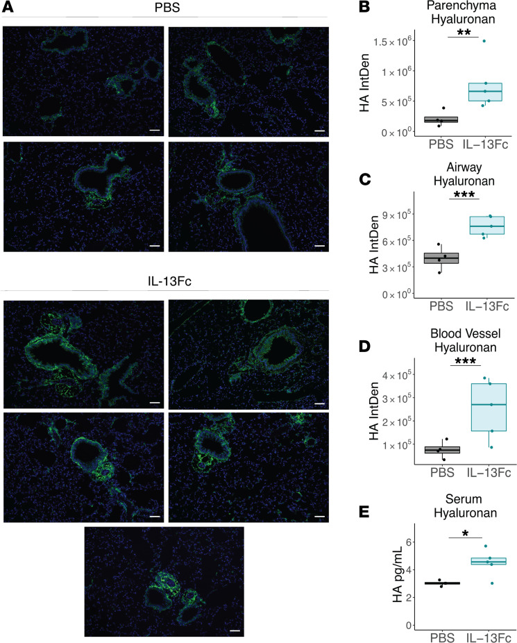
Immune dysregulation is characteristic of the more severe stages of SARS-CoV-2 infection. Understanding the mechanisms by which the immune system contributes to COVID-19 severity may open new avenues to treatment. Here, we report that elevated IL-13 was associated with the need for mechanical ventilation in 2 independent patient cohorts. In addition, patients who acquired COVID-19 while prescribed Dupilumab, a mAb that blocks IL-13 and IL-4 signaling, had less severe disease. In SARS-CoV-2-infected mice, IL-13 neutralization reduced death and disease severity without affecting viral load, demonstrating an immunopathogenic role for this cytokine. Following anti-IL-13 treatment in infected mice, hyaluronan synthase 1 (Has1) was the most downregulated gene, and accumulation of the hyaluronan (HA) polysaccharide was decreased in the lung. In patients with COVID-19, HA was increased in the lungs and plasma. Blockade of the HA receptor, CD44, reduced mortality in infected mice, supporting the importance of HA as a pathogenic mediator. Finally, HA was directly induced in the lungs of mice by administration of IL-13, indicating a new role for IL-13 in lung disease. Understanding the role of IL-13 and HA has important implications for therapy of COVID-19 and, potentially, other pulmonary diseases. IL-13 levels were elevated in patients with severe COVID-19. In a mouse model of the disease, IL-13 neutralization reduced the disease and decreased lung HA deposition. Administration of IL-13-induced HA in the lung. Blockade of the HA receptor CD44 prevented mortality, highlighting a potentially novel mechanism for IL-13-mediated HA synthesis in pulmonary pathology.

Keywords: COVID-19; Cytokines; Immunology; Innate immunity; Th2 response.


Figures

Figure 1
Figure 1. Type 2 immune response in patients with severe COVID-19 disease.
(AE) Cytokines were measured in plasma from 26 outpatients and 152 inpatients with COVID-19 infection at the University of Virginia Hospital using a 48-plex cytokine array. (A) Heatmap of plasma cytokines, supplemental oxygen requirement and nasopharyngeal viral load, with rows ordered by patient status (outpatient (OP) versus inpatient (IP)) and columns by cytokine PC 1 which included IL-13 (Supplemental Table 2). (B) Scatterplot comparing PC 1 and 2 from PCA of the plasma cytokines (orange inpatients and blue outpatients). (C) Plasma IL-13 levels in patients with COVID-19 who were or were not diagnosed with COVID-19 or (D) did or did not require mechanical ventilation (Wilcox test). Data shown by box-and-whisker plots representing the median, interquartile range (box), upper and lower quartiles (whiskers), and outliers as points falling outside the bounds of the upper and lower quartiles. (E) Kaplan-Meier survival analysis of the relationship between IL-13 level and mechanical ventilation. Comparison made to lowest IL-13 quantile (Cox proportional hazards test adjusted for age, sex, and comorbidities). (F) Proportion of patients with COVID-19 requiring mechanical ventilation stratified by IL-13 plasma cytokine levels (χ2 analysis). (G) ROC curve with AUC plotted from: IL-13 alone (blue); IL-13 and IL-6 (red); or IL-13, IL-6, IL-8, and MIP-1b (black). (H) IL-13 levels in 19 nonsevere and 26 severe (requiring supplemental oxygen) patients with COVID-19 from Virginia Commonwealth University Hospital (Wilcox test). *P < 0.05; **P < 0.005; ****P < 0.0001.
Figure 2
Figure 2. Type 2 immune response in lungs of mice following infection with SARS-CoV-2.
Ten-week-old male mice (Tg K18-hACE2 2Prlmn) were infected with 5 × 10³ PFU of SARS-CoV-2 and lung tissue examined on day 5 after infection by RNA-Seq and IHC. (A) Type 2 gene expression in the lungs of infected versus uninfected mice (heatmap of normalized values of manually curated list of type 2 immune pathway genes). (B) IHC of the type 2 immunity proteins RELMα and Ym1 in the lungs of infected and uninfected mice. (C) Quantification of IHC scoring for RELMα and Ym1 (mixed effect model). Scale bar: 70 μm. n = 5 mice/group. **P < 0.005.
Figure 3
Figure 3. IL-13 neutralization protects from severe COVID-19 in K18-hACE2 mice and dupilumab use is associated with protection in humans.
Mice were infected on day 0 with 5 × 10³ PFU of SARS-CoV-2 and administered 150 μg of anti–IL-13 or an IgG isotype control antibody i.p. on days 0, 2, and 4. (A) Clinical scores of illness severity on days 1–7 after infection. Clinical scoring was measured by weight loss (scores 0–5), posture and appearance of fur (piloerection) (scores 0–2), activity (scores 0–3), and eye closure (scores 0–2). (B) Weight loss on days 1–7 after infection. (A and B Student’s t test). (C) Kaplan-Meier survival analysis in mice. (D) Kaplan-Meier curve generated from data obtained from TriNetX: 1:1 matching based on 81 patients who had been prescribed Dupilumab independently of their COVID-19 diagnosis. (E) Quantification of intensity of staining for parenchyma and epithelial RELMα following IL-13 neutralization (log-transformed, mixed effect model). (F) IHC of lung tissue stained for RELMα (yellow) in parenchyma or airway, and DAPI (blue). (n = 5 mice/group; (AC) combined from 3 independently conducted experiments).
Figure 4
Figure 4. HA and COVID-19 disease.
Mice received i.p. injections of anti–IL-13 on days 0 and 2 after infection, were euthanized on day 5, and lung tissue was split and placed either into trizol tissue reagent for RNA analysis or formaldehyde for paraffin embedding and IHC. (A) Gene expression in mouse lung of Has1, the HA receptor Cd44, and Hyaluronidase 2 (Has2) of infected mice with anti–IL-13, isotype control antibody and uninfected controls (Tukey’s HSD). (B) Staining of HA in mouse lung (with HABP); Scale bar: 70μm. Rectangle indicates area magnified in image to left; Scale bar: 20μm. (C) Quantification of HA deposition in tissue following infection and neutralization of IL-13 (mixed effect model; combined 2 experiments). (D) Hyaluronan was measured in the plasma of COVID-19–negative controls and in patients with COVID-19 that did or did not require supplemental oxygen. Postmortem lung samples were obtained from fatal COVID-19 cases and control tissue from COVID-19 negative deaths. (E) Quantification of HA deposition in fatal COVID-19 disease (n = 11) and controls (n = 8) (log-transformed, mixed effect model) using HABP. (F) Representative images of staining for HA (with HABP) from human lung samples; Scale bar: 70μm. Rectangle indicates area magnified in image below; Scale bar: 20μm. Mice were administered anti-CD44 or IgG2 isotype control on each of day 1–4 after infection. (G) Kaplan-Meier survival curve (log-rank) and (H) Clinical scores for mice (Student’s t test); combined 2 independent experiments. Hyaluronan-binding protein, HABP. n = 5 mice/group. *P < 0.05; **P < 0.005; ***P < 0.0005.
Figure 5
Figure 5. IL-13 administration promotes HA accumulation in mice.
Uninfected mice were administered IL-13 Fc (n = 5) or PBS (n = 4) on days 0 and 2 and lung tissue and serum was collected 24 hours later. Lung tissue was sectioned and stained for HABP. (A) Representative images from each mouse. Quantification of HAPB staining in the (B) parenchyma, (C) airway, and (D) blood vessels for quantification of HA deposition in the tissue. (BD; mixed effect model). (E) Hyaluronan was measured in the serum by ELISA (Student’s t test). Hyaluronan-binding protein, HABP. Scale bar: 70μm. *P < 0.05; **P < 0.01; ***P < 0.001.

Update of

  • IL-13 is a driver of COVID-19 severity.
    Donlan AN, Sutherland TE, Marie C, Preissner S, Bradley BT, Carpenter RM, Sturek JM, Ma JZ, Moreau GB, Donowitz JR, Buck GA, Serrano MG, Burgess SL, Abhyankar MM, Mura C, Bourne PE, Preissner R, Young MK, Lyons GR, Loomba JJ, Ratcliffe SJ, Poulter MD, Mathers AJ, Day A, Mann BJ, Allen JE, Petri WA Jr. Donlan AN, et al. medRxiv [Preprint]. 2021 Mar 1:2020.06.18.20134353. doi: 10.1101/2020.06.18.20134353. medRxiv. 2021. Update in: JCI Insight. 2021 Aug 9;6(15):150107. doi: 10.1172/jci.insight.150107. PMID: 33688686 Free PMC article. Updated. Preprint.

References

    1. Cantuti-Castelvetri L, et al. Neuropilin-1 facilitates SARS-CoV-2 cell entry and infectivity. Science. 2020;370(6518):856–860. doi: 10.1126/science.abd2985. - DOI - PMC - PubMed
    1. Hoffmann M, et al. SARS-CoV-2 cell entry depends on ACE2 and TMPRSS2 and is blocked by a clinically proven protease inhibitor. Cell. 2020;181(2):271–280. doi: 10.1016/j.cell.2020.02.052. - DOI - PMC - PubMed
    1. Pedersen SF, Ho YC. SARS-CoV-2: a storm is raging. J Clin Invest. 2020;130(5):2202–2205. doi: 10.1172/JCI137647. - DOI - PMC - PubMed
    1. Mangalmurti N, Hunter CA. Cytokine storms: understanding COVID-19. Immunity. 2020;53(1):19–25. doi: 10.1016/j.immuni.2020.06.017. - DOI - PMC - PubMed
    1. Tisoncik JR, et al. Into the eye of the cytokine storm. Microbiol Mol Biol Rev. 2012;76(1):16–32. doi: 10.1128/MMBR.05015-11. - DOI - PMC - PubMed