Review
. 2021 Oct:344:113795.
doi: 10.1016/j.expneurol.2021.113795. Epub 2021 Jun 26.

Neurotoxicity of methamphetamine: Main effects and mechanisms

Affiliations
Review

Neurotoxicity of methamphetamine: Main effects and mechanisms

Subramaniam Jayanthi et al. Exp Neurol. 2021 Oct.

Abstract

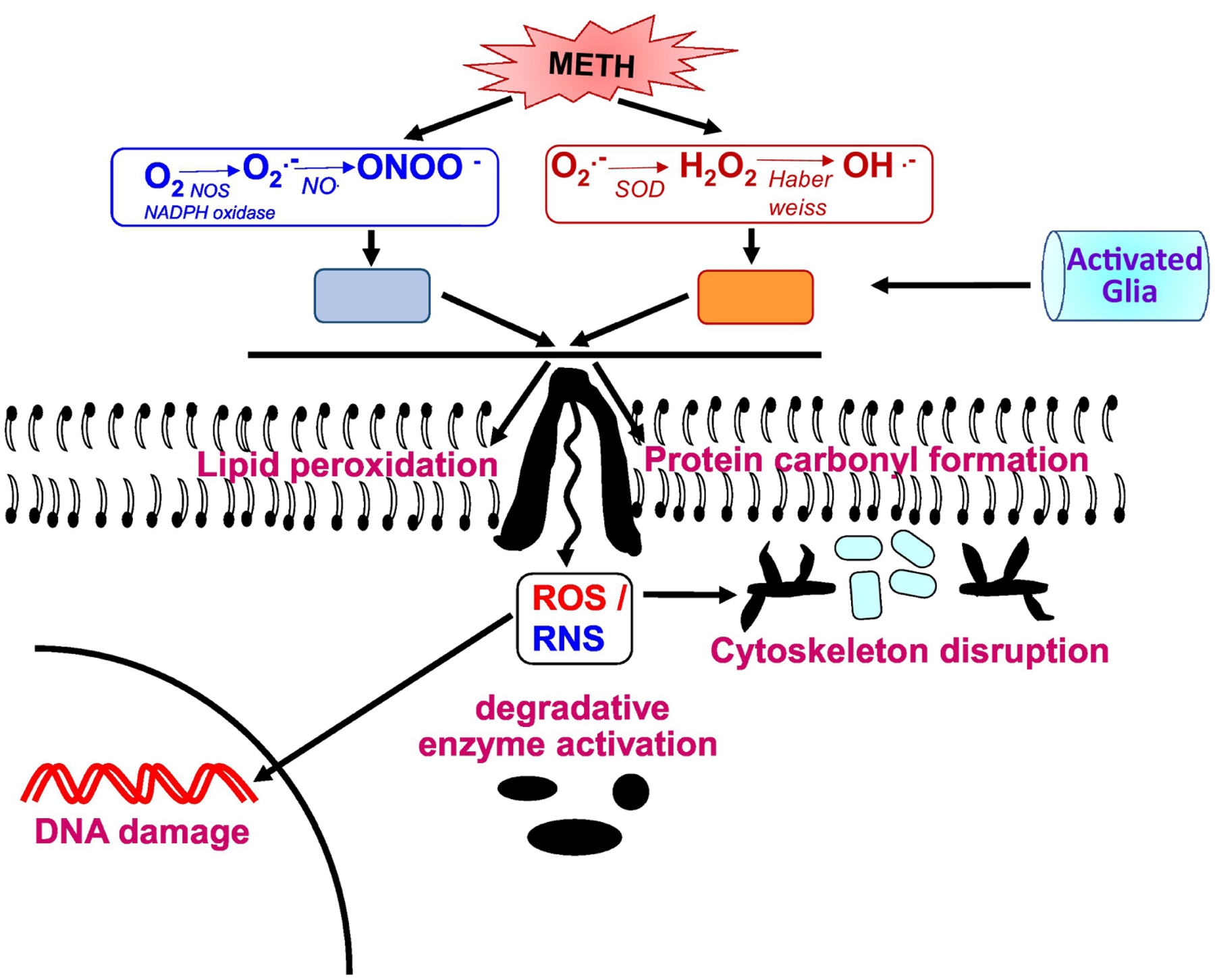
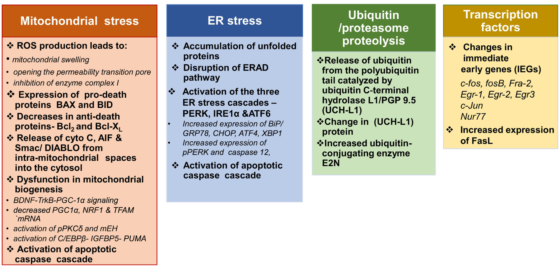
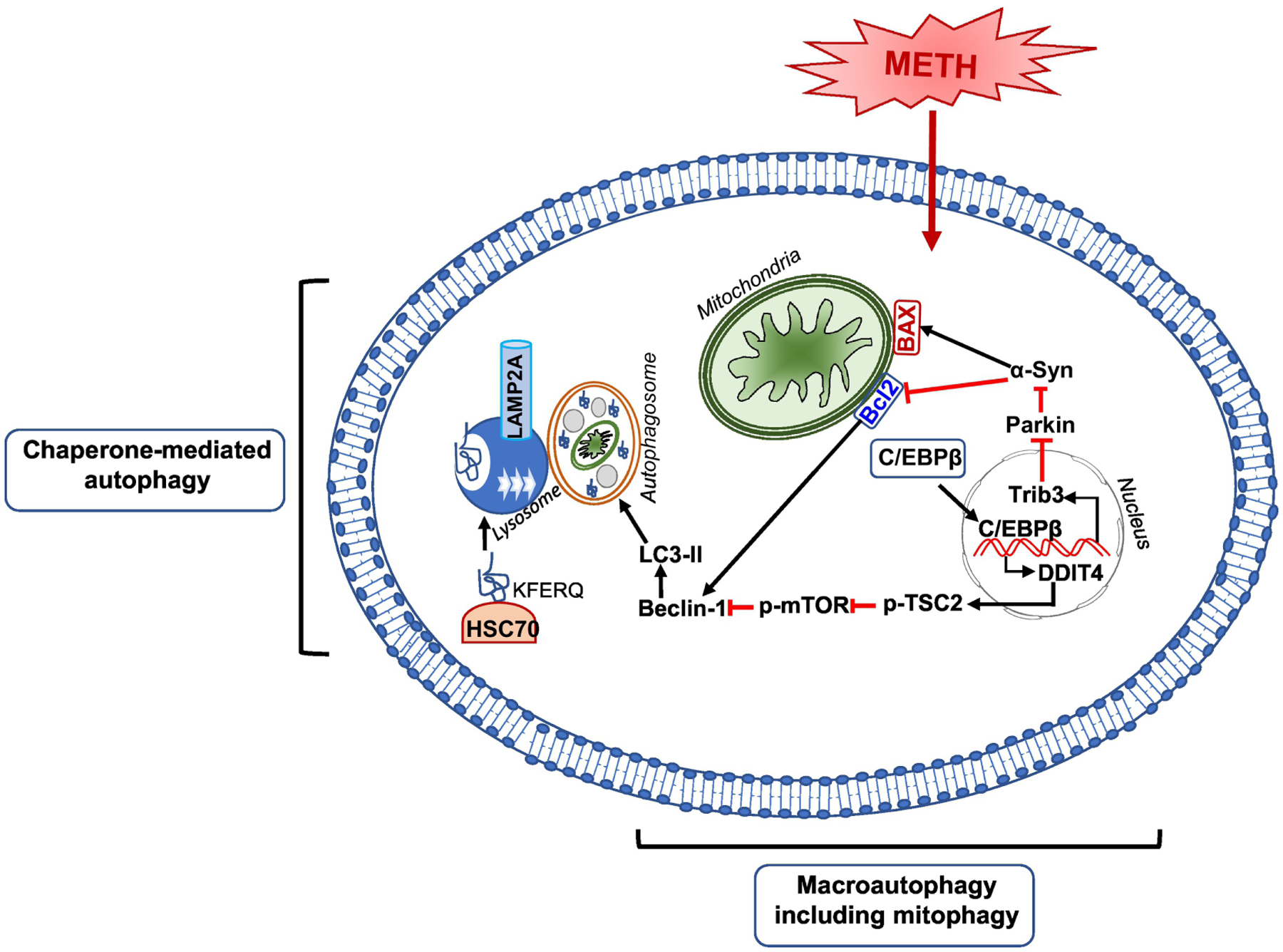
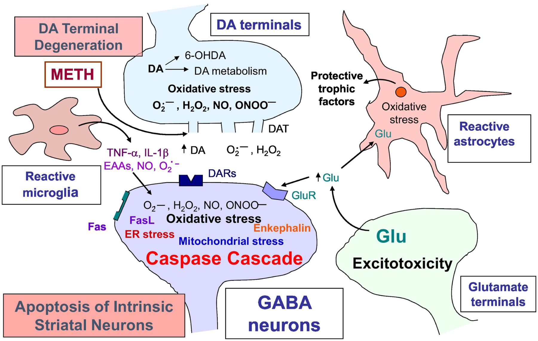
Methamphetamine (METH) is an illicit psychostimulant that is abused throughout the world. METH addiction is also a major public health concern and the abuse of large doses of the drug is often associated with serious neuropsychiatric consequences that may include agitation, anxiety, hallucinations, paranoia, and psychosis. Some human methamphetamine users can also suffer from attention, memory, and executive deficits. METH-associated neurological and psychiatric complications might be related, in part, to METH-induced neurotoxic effects. Those include altered dopaminergic and serotonergic functions, neuronal apoptosis, astrocytosis, and microgliosis. Here we have endeavored to discuss some of the main effects of the drug and have presented the evidence supporting certain of the molecular and cellular bases of METH neurotoxicity. The accumulated evidence suggests the involvement of transcription factors, activation of dealth pathways that emanate from mitochondria and endoplasmic reticulum (ER), and a role for neuroinflammatory mechanisms. Understanding the molecular processes involved in METH induced neurotoxicity should help in developing better therapeutic approaches that might also serve to attenuate or block the biological consequences of use of large doses of the drug by some humans who meet criteria for METH use disorder.

Keywords: Autophagy; Bcl2; Cell death; ER stress; Mitochondrial cascade; Neuroinflammation; Transcription factors.


Conflict of interest statement

Conflict of Interest

The authors declare that they have no conflict of interest.

Figures

Figure 1.
Figure 1.. Methamphetamine neurotoxicity- ROS and neuroinflammation.
METH augments ROS and RNS levels that eventually lead to oxidative stress state. ROS in excessive concentrations can cause cellular damage to DNA, lipid membranes, proteins and other macromolecules. The end products of lipid peroxidation and protein carbonyl oxidation cause cytoskeleton disruption and DNA damage, such as double-strand DNA breaks. Moreover, the reactive species (ROS and RNS) triggers signaling pathways that lead to the over-activation of the major glial inflammatory characters: microglia and astrocytes. These glial cells are the mediators of neuroinflammatory response and ultimately leads to neurodegeneration.
Figure 2.
Figure 2.. Methamphetamine neurotoxicity-cell death mechanisms.
Scheme summarizes insights into the various molecular and functional connections between the different METH-induced neuronal cell death mechanisms.
Figure 3.
Figure 3.. Methamphetamine neurotoxicity-autophagy.
Schematic figure shows the signaling pathways that may be involved in METH-induced autophagy. METH is shown to facilitate both chaperone-mediated autophagy (CMA) and macro-autophagy. In both autophagic mechanisms, Beclin1 is the vital protein that associates with Bcl2 and promotes autophagy. METH-induced macro-autophagy is mediated via C/EBPβ by induction of C/EBPβ/DDIT4 /TSC2/mTOR signal axis or induction of C/EBPβ/Trib3 /Parkin/α-Syn signal axis. For the METH-induced CMA, The KFERQ-like motif of a cargo protein is detected by the chaperone, HSC70. This complex binds to the lysosomal membrane protein lysosome-associated membrane protein type 2A (LAMP2A). Then, the assembled LAMP2A is translocated through the lysosomal membrane. Once inside the lysosomal lumen, the substrate protein is rapidly degraded by lysosomal proteases and the hsc70 chaperone complex is released from the lysosome and ready to bind to another substrate protein for CMA.
Figure 4.
Figure 4.. Possible mechanisms of neurotoxicity produced by methamphetamine.
The depicted scheme provides a well- studied mechanistic outline in METH neurotoxicity; however, it mainly refers to the striatal synapses. Large doses of METH causes degeneration of DA terminals characterized by excessive DA release from the storage vesicles and causes perturbation in DA metabolism. Extracellular DA auto-oxidizes to produce reactive oxygen (O2.-, H2O2) and nitrogen (NO, ONOO-) species (ROS/RNS). METH exposure also causes increased activity of microglia and astrocytes. Reactive gliosis is associated with increased production of pro-inflammatory cytokines (TNFα; IL-1β) and ROS/RNS. This process initiates oxidative-stress mediated neuronal apoptotic cascade that includes mitochondrial stress, endoplasmic reticulum (ER) stress, ubiquitin-proteasome proteolysis, and neuroinflammation. In addition to affecting DA transmission, METH also exerts excitotoxicity via glutamate release that binds to glutamate receptors (GluR), triggers calcium influx, produces RNS and further oxidative stress-induced damage.

References

    1. Achat-Mendes C, Ali SF, Itzhak Y (2005) Differential effects of amphetamines-induced neurotoxicity on appetitive and aversive pavlovian conditioning in mice. Neuropsychopharmacology 30:1128–1137. - PubMed
    1. Allagnat F, Fukaya M, Nogueira TC, Delaroche D, Welsh N, Marselli L, Marchetti P, Haefliger JA, Eizirik DL, Cardozo AK (2012) C/EBP homologous protein contributes to cytokine-induced pro-inflammatory responses and apoptosis in β-cells. Cell Death Differ 19:1836–1846. - PMC - PubMed
    1. Ando K, Johanson CE, Seiden LS, Schuster CR (1985) Sensitivity changes to dopaminergic agents in fine moto control of rhesus monkeys after repeated methamphetamine administration. Pharmacology Biochemistry and Behavior 22:737–743. - PubMed
    1. Ares-Santos S, Granado N, Moratalla R (2013) The role of dopamine receptors in the neurotoxicity of methamphetamine. J Intern Med 273:437–453. - PubMed
    1. Ares-Santos S, Granado N, Oliva I, Martin ED, Colado MI, Moratalla R (2012) Dopamine D (1) receptor deletion strongly reduces neurotoxic effects of methamphetamine. Neurobiol Dis 45:810–820. - PubMed

Publication types

MeSH terms

Substances