Comparative Study
. 2021:20:100120.
doi: 10.1016/j.mcpro.2021.100120. Epub 2021 Jun 27.

Comparative Host Interactomes of the SARS-CoV-2 Nonstructural Protein 3 and Human Coronavirus Homologs

Affiliations
Comparative Study

Comparative Host Interactomes of the SARS-CoV-2 Nonstructural Protein 3 and Human Coronavirus Homologs

Katherine M Almasy et al. Mol Cell Proteomics. 2021.

Abstract

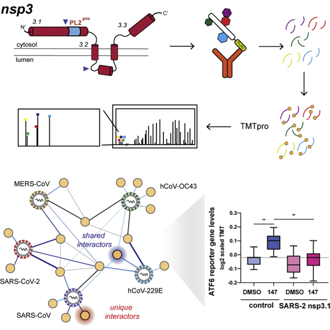
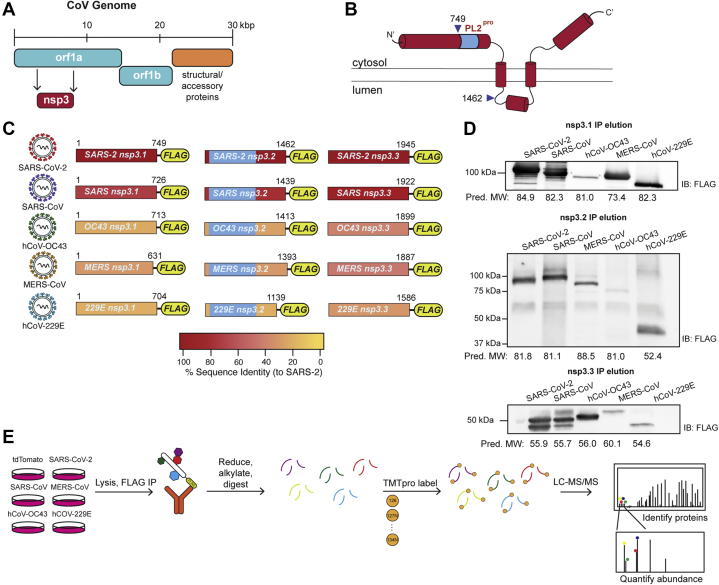
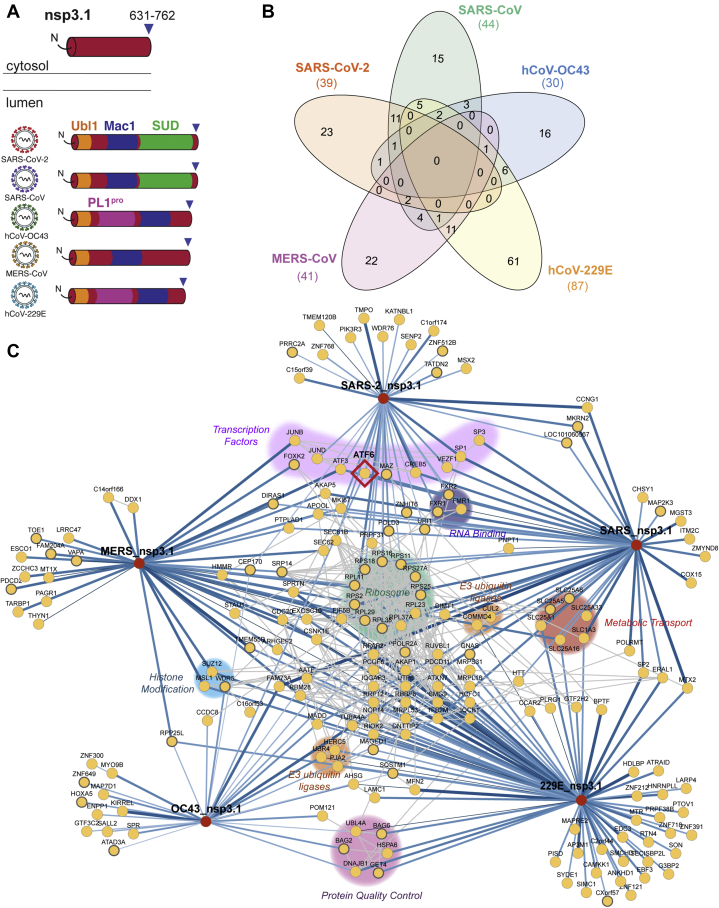
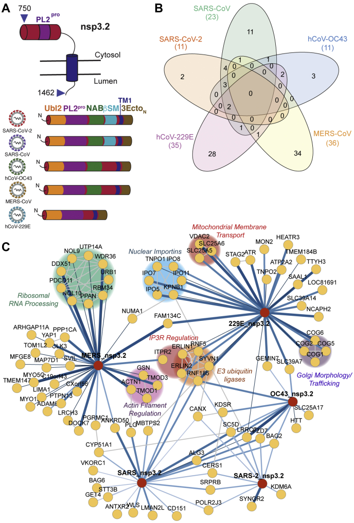
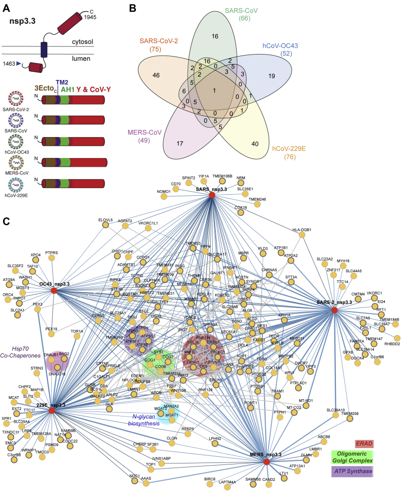
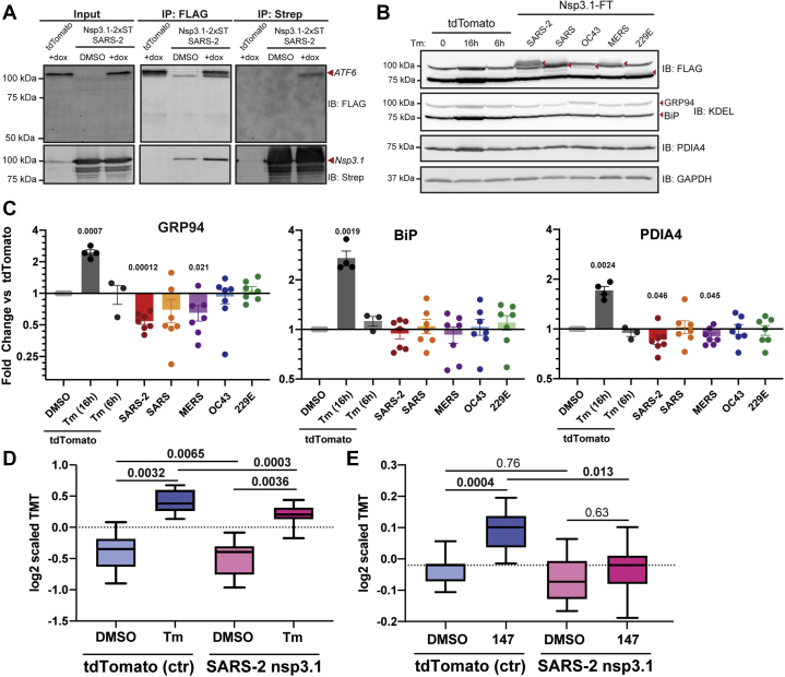
Human coronaviruses have become an increasing threat to global health; three highly pathogenic strains have emerged since the early 2000s, including most recently SARS-CoV-2, the cause of COVID-19. A better understanding of the molecular mechanisms of coronavirus pathogenesis is needed, including how these highly virulent strains differ from those that cause milder, common-cold-like disease. While significant progress has been made in understanding how SARS-CoV-2 proteins interact with the host cell, nonstructural protein 3 (nsp3) has largely been omitted from the analyses. Nsp3 is a viral protease with important roles in viral protein biogenesis, replication complex formation, and modulation of host ubiquitinylation and ISGylation. Herein, we use affinity purification-mass spectrometry to study the host-viral protein-protein interactome of nsp3 from five coronavirus strains: pathogenic strains SARS-CoV-2, SARS-CoV, and MERS-CoV; and endemic common-cold strains hCoV-229E and hCoV-OC43. We divide each nsp3 into three fragments and use tandem mass tag technology to directly compare the interactors across the five strains for each fragment. We find that few interactors are common across all variants for a particular fragment, but we identify shared patterns between select variants, such as ribosomal proteins enriched in the N-terminal fragment (nsp3.1) data set for SARS-CoV-2 and SARS-CoV. We also identify unique biological processes enriched for individual homologs, for instance, nuclear protein import for the middle fragment of hCoV-229E, as well as ribosome biogenesis of the MERS nsp3.2 homolog. Lastly, we further investigate the interaction of the SARS-CoV-2 nsp3 N-terminal fragment with ATF6, a regulator of the unfolded protein response. We show that SARS-CoV-2 nsp3.1 directly binds to ATF6 and can suppress the ATF6 stress response. Characterizing the host interactions of nsp3 widens our understanding of how coronaviruses co-opt cellular pathways and presents new avenues for host-targeted antiviral therapeutics.

Keywords: COVID-19; activating transcription factor 6; affinity purification-mass spectrometry; nsp3; tandem mass tags; unfolded protein response.


Conflict of interest statement

Conflict of interest The authors declare that they have no conflict of interest.

Figures

None
Graphical abstract
Fig. 1
Fig. 1
Design and expression of CoV nsp3 truncations for affinity purification–mass spectrometry (AP-MS).A, coronavirus (CoV) genome schematic indicating the regions encoding orf1a, orf1ab, and structural/accessory proteins. Nsp3 is encoded within orf1a. B, general schematic of SARS-CoV-2 nsp3 protein topology. Nsp3 has two single-span transmembrane regions anchoring the protein in the ER membrane, with a small luminal domain and both N and C terminal regions in the cytosol. The conserved papain-like protease (PL2pro) domain is on the N-terminal cytosolic portion. For this study, the nsp3 protein was truncated into three fragments: nsp3.1 (1–749), nsp3.2 (750–1462), and nsp3.3 (1463–1945), numbering corresponding to SARS-CoV-2 nsp3. C, the nsp3 truncations for homologs from all five human coronaviruses used in this study. All fragments contain a C-terminal FLAG tag for affinity purification. Percent sequence identity compared with SARS-CoV-2 is indicated. The PL2pro domain in nsp3.2 homologs is highlighted in light blue. D, western blotting of immunopurified nsp3.1, nsp3.2, and nsp3.3 homologs after transient transfection in HEK293T cells. Predicted MW of proteins is indicated below. E, AP-MS workflow to identify virus–host protein interactions of nsp3 fragment homologs. HEK293T cells were transfected with corresponding homologs and lysates were immunopurified using anti-FLAG beads to enrich for viral proteins in complex with host interactors. Proteins were reduced, alkylated, and digested with trypsin/LysC. Peptides were then labeled with tandem mass tags (TMTpro) and analyzed by tandem mass spectrometry (LC-MS/MS) to both identify and quantify host interactors.
Fig. 2
Fig. 2
Identification of CoV nsp3.1 host interactors.A, schematic of nsp3.1 topology for all five CoV homologs. Nsp3.1 is a cytosolic fragment, comprising residues from 1 to 631–762. All fragment homologs contain a ubiquitin-like domain (Ubl1, yellow) and a conserved macrodomain (Mac1, blue). SARS-CoV-2 and SARS-CoV contain a SARS-unique domain (SUD, green), while hCoV-OC43 and hCoV-229E contain a papain-like protease domain (PL1pro, pink). B, Venn diagram showing the number of unique and shared host interactors amongst all five CoV nsp3.1 homologs. Total interactors for each homolog are shown in parentheses. C, network plot of virus–host interactors. Individual nsp3.1 homologs are shown as red circles, while host interactors are shown as yellow circles. Blue lines indicate virus–host protein interactions, where line width and shade are wider/darker for more highly enriched interactions. Gray lines indicate known host–host protein interactions from the STRING database. Notable clusters of host proteins are highlighted. The transcription factor ATF6 (red diamond) interacts with SARS-CoV-2, SARS-CoV, and hCoV-OC43 and was subjected to later functional follow-up (Fig. 5).
Fig. 3
Fig. 3
Comparative interactomics of CoV nsp3.2 homologs.A, schematic of nsp3.2 protein topology for all five CoV homologs. Nsp3.2 contains both cytosolic, transmembrane, and luminal regions. All homologs contain a ubiquitin-like domain (Ubl2, yellow), a papain-like protease domain (PL2pro, purple), a transmembrane region (TM1, blue), and the N-terminal portion of the ectodomain (3EctoN, brown). Betacoronavirus homologs also contain a nucleic acid binding domain (NAB, green), while SARS strains also have a betacoronavirus-specific marker (βSM, light blue). B, Venn diagram showing the number of unique and shared host interactors amongst all five CoV nsp3.2 homologs. Total interactors for each homolog are shown in parentheses. C, network plot of virus–host interactors. Individual nsp3.2 homologs are shown as red circles, while host interactors are shown as yellow circles. Blue lines indicate virus–host protein interactions, where line width and shade are wider/darker for more highly enriched interactions. Gray lines indicate known host–host protein interactions from the STRING database. Notable clusters of host proteins are highlighted.
Fig. 4
Fig. 4
Identification of CoV nsp3.3 host interactors.A, schematic of nsp3.3 protein topology for all five CoV homologs. Nsp3.3 contains cytosolic, transmembrane, and luminal regions. All homologs contain the C-terminal portion of the ectodomain (3EctoC, brown), the second transmembrane region (TM2, blue), a likely amphipathic helix (AH1, green), and a Y&CoV-Y domain (red). B, Venn diagram showing the number of unique and shared host interactors among all five CoV nsp3.3 homologs. Total interactors for each homolog are shown in parentheses. C, network plot of virus–host interactors. Individual nsp3.3 homologs are shown as red circles, while host interactors are shown as yellow circles. Blue lines indicate virus–host protein interactions, where line width and shade are wider/darker for more highly enriched interactions. Gray lines indicate known host–host protein interactions from the STRING database. Notable clusters of host proteins are highlighted.
Fig. 5
Fig. 5
SARS-CoV-2 nsp3.1 interacts with ATF6 and suppresses the ATF6 branch of the unfolded protein response (UPR).A, representative coimmunopurification (IP) western blots of 3xFT-ATF6 cells transfected with SARS-CoV-2 nsp3.1-2xST or tdTomato (control), lysed, and immunopurified for either FLAG or 2xStrepTag. Cells were treated with 100 nM doxycycline to induce 3xFT-ATF6 expression 24 h preharvest. Input and IP elution blots were probed with both anti-FLAG and anti-StrepTag antibodies. n = 3. B, representative western blots of HEK293T cells transfected with nsp3.1-FT homologs or tdTomato (control), lysed, and probed for FLAG, ATF6 branch markers (GRP94, BiP, PDIA4), and GAPDH as a loading control. Control cells were either treated with DMSO or 6 μg/ml Tunicamycin (Tm) at 6 or 16 h preharvest to induce an ER stress response. TdTomato 16 h preharvest, n = 4; TdTomato 6 h preharvest, n = 3; all others, n = 7. C, quantification of western blots shown in (B). Error bars indicate average ±SEM. Paired Student’s t-tests were used to test for significance between samples and tdTomato+DMSO control, with p-values <0.05 shown. D, box-and-whisker plots of ATF6-regulated protein abundance measured by quantitative proteomics. HEK293T cells were transfected with tdTomato (control) or SARS-CoV-2 nsp3.1-FT and treated with DMSO or 6 μg/ml tunicamycin for 16 h preharvest. Shown are the distribution of scaled log2 TMT intensities for ATF6-regulated proteins based on published genesets (50). A one-way ANOVA with Geisser–Greenhouse correction and post-hoc Tukey’s multiple comparison test was used to determine significance. Adjusted p-values are shown. n = 4 biological replicates in a single mass spectrometry run. E, box-and-whisker plots of ATF6-regulated protein abundance measured by quantitative proteomics. HEK293T cells were transfected with tdTomato (control) or SARS-CoV-2 nsp3.1-FT and treated with DMSO or 10 μM 147 for 16 h preharvest. Shown are the distribution of scaled log2 TMT intensities for ATF6-regulated proteins based on published genesets (50). A one-way ANOVA with Geisser–Greenhouse correction and post-hoc Tukey’s multiple comparison test was used to determine significance. Adjusted p-values are shown. n = 3 biological replicates in a single mass spectrometry run.

Update of

References

    1. Ksiazek T.G., Erdman D., Goldsmith C.S., Zaki S.R., Peret T., Emery S., Tong S., Urbani C., Comer J.A., Lim W., Rollin P.E., Dowell S.F., Ling A.-E., Humphrey C.D., Shieh W.-J. A novel coronavirus associated with severe acute respiratory syndrome. N. Engl. J. Med. 2003;348:1953–1966. - PubMed
    1. Drosten C., Günther S., Preiser W., van der Werf S., Brodt H.-R., Becker S., Rabenau H., Panning M., Kolesnikova L., Fouchier R.A.M., Berger A., Burguière A.-M., Cinatl J., Eickmann M., Escriou N. Identification of a novel coronavirus in patients with severe acute respiratory syndrome. N. Engl. J. Med. 2003;348:1967–1976. - PubMed
    1. Zaki A.M., van Boheemen S., Bestebroer T.M., Osterhaus A.D.M.E., Fouchier R.A.M. Isolation of a novel coronavirus from a man with pneumonia in Saudi Arabia. N. Engl. J. Med. 2012;367:1814–1820. - PubMed
    1. Wu F., Zhao S., Yu B., Chen Y.M., Wang W., Song Z.G., Hu Y., Tao Z.W., Tian J.H., Pei Y.Y., Yuan M.L., Zhang Y.L., Dai F.H., Liu Y., Wang Q.M. A new coronavirus associated with human respiratory disease in China. Nature. 2020;579:265–269. - PMC - PubMed
    1. Zhou P., Yang X.L., Wang X.G., Hu B., Zhang L., Zhang W., Si H.R., Zhu Y., Li B., Huang C.L., Chen H.D., Chen J., Luo Y., Guo H., Jiang R.D. A pneumonia outbreak associated with a new coronavirus of probable bat origin. Nature. 2020;579:270–273. - PMC - PubMed

Publication types

MeSH terms

Substances