. 2021 Jun 29;16(1):43.
doi: 10.1186/s13024-021-00466-z.

AMPK hyperactivation promotes dendrite retraction, synaptic loss, and neuronal dysfunction in glaucoma

Affiliations

AMPK hyperactivation promotes dendrite retraction, synaptic loss, and neuronal dysfunction in glaucoma

Nicolas Belforte et al. Mol Neurodegener. .

Abstract

Background: The maintenance of complex dendritic arbors and synaptic transmission are processes that require a substantial amount of energy. Bioenergetic decline is a prominent feature of chronic neurodegenerative diseases, yet the signaling mechanisms that link energy stress with neuronal dysfunction are poorly understood. Recent work has implicated energy deficits in glaucoma, and retinal ganglion cell (RGC) dendritic pathology and synapse disassembly are key features of ocular hypertension damage.

Results: We show that adenosine monophosphate-activated protein kinase (AMPK), a conserved energy biosensor, is strongly activated in RGC from mice with ocular hypertension and patients with primary open angle glaucoma. Our data demonstrate that AMPK triggers RGC dendrite retraction and synapse elimination. We show that the harmful effect of AMPK is exerted through inhibition of the mammalian target of rapamycin complex 1 (mTORC1). Attenuation of AMPK activity restores mTORC1 function and rescues dendrites and synaptic contacts. Strikingly, AMPK depletion promotes recovery of light-evoked retinal responses, improves axonal transport, and extends RGC survival.

Conclusions: This study identifies AMPK as a critical nexus between bioenergetic decline and RGC dysfunction during pressure-induced stress, and highlights the importance of targeting energy homeostasis in glaucoma and other neurodegenerative diseases.

Keywords: Adenosine monophosphate-activated protein kinase; Glaucoma; Mammalian target of rapamycin; Metabolic stress; Neurodegeneration.


Conflict of interest statement

The authors declare that they have no competing interests.

Figures
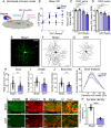
Fig. 1
Fig. 1
Ocular hypertension promotes early RGC dendrite retraction and synapse loss. A Schematic of magnetic microbeads injection into the mouse anterior chamber to induce ocular hypertension (OHT). B This procedure results in gradual increase of intraocular pressure (IOP) leading to RGC soma and axon loss (ANOVA with Tukey’s multiple comparison post-hoc test, ***p < 0.001, N = 10–14 mice/group). C, D Quantitative analysis demonstrates a statistically significant loss of RGC soma and axons at 3 weeks after microbeads injection (ANOVA with Tukey’s multiple comparison post-hoc test, n.s. = not significant, ***p < 0.001, N = 5–14 mice per group). (E) YFP-positive RGC with a clearly identifiable axon (arrowhead) were selected for dendritic arbor imaging and 3D reconstruction. F, G Representative examples of RGC dendritic arbors from non-injured (sham) and glaucomatous retinas at 2 weeks after induction of OHT. H-K Quantitative analysis of dendritic parameters reveals a substantial reduction in dendritic area, length, number of branches, and complexity (Sholl analysis) in OHT retinas (blue) relative to sham controls (white) (Student’s t-test, **p < 0.01, ***p < 0.001, N = 6 mice/group, n = 40–51 RGC/group Table 1). L-N Glutamatergic synapses are visualized in the inner plexiform layer (IPL) on retinal cross-sections using immunolabeling against PSD95 (green) and VGLUT1 (red), post- and pre-synaptic markers, respectively. OHT injury (2 weeks) induces a pronounced loss of both VGLUT1 and PSD95 expression in the IPL. O High magnification of inset in (N) shows VGLUT1 and PSD95 puncta in the IPL at the level of the OFF sublamina in OHT and sham retinas. P Quantitative analysis of pre- and postsynaptic co-localized voxels, which measured the three-dimensional volume occupied by both VGLUT1 and PSD95 in the IPL, confirms that OHT promotes a striking loss of synapses (Student’s t-test, ***p < 0.001, N = 4 mice/group). Values are expressed as the mean ± S.E.M
Fig. 2
Fig. 2
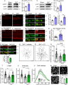
Glaucoma-induced energy stress triggers AMPK activation. A, B Western blot and densitometry analysis of retinal homogenates demonstrate a substantial increase in active AMPK (pAMPKThr172), a readout of metabolic stress, as early as 1 week after induction of OHT (Student’s t-test, * = p<0.05, N = 5 mice/group). The lower panel is the same blot probed with an antibody against total AMPK and β-actin for normalization. C, D Western blot and densitometry analysis of retinal homogenates show increased LKB1 activity (pLKB1) in glaucomatous retinas (Student’s t-test, ** = p<0.01, N = 7–9 mice/group). The lower panel is the same blot probed with an antibody against total LKB1 and β-actin for normalization. E, F Immunohistochemical analysis of mouse retinal cross sections with an antibody against pAMPKThr172, reveals robust AMPK activity in retinal cells subjected to OHT. G, H Co-labeling with antibodies against pAMPKThr172 and the RGC-specific marker Brn3a demonstrates AMPK hyperactivity in RGC. I, J Quantification of the number of pAMPKThr172-positive RGC as well as epifluorescence intensity per neuron confirms a significant increase in AMPK activity (Student’s t-test, ** = p<0.01, N = 5 mice/group, n = 50 RGC/group). K, L pAMPKThr172 retinal immunostaining of primary open angle glaucoma patients and age-matched controls (Table 2) reveals increased AMPK function. M, N Co-labeling with anti-pAMPKThr172 and RBPMS, a selective marker for RGC, confirms AMPK overactivation in glaucomatous RGC. O Quantification of epifluorescence intensity in pAMPKThr172-positive RGC demonstrates a two-fold increase in AMPK activity in human glaucomatous retinas relative to age-matched controls (Student’s t-test, ** = p<0.01, glaucoma: N = 27 retinas/group, n = 100 RGC/group; controls: N = 15 retinas/group, n = 100 RGC/group). P Administration of compound C (CC), an inhibitor of AMPK, results a significant decrease in AMPK activity compared to vehicle-treated eyes. (Student’s t-test, *** = p<0.001, N = 5 mice/group). Q, R Representative examples of dendritic arbors from OHT retinas treated with vehicle or CC, an inhibitor of AMPK, visualized at 2 weeks after microbeads injection. S-V Quantitative analysis of dendritic parameters reveals that CC-treated neurons had longer dendrites and markedly larger and more complex arbors than vehicle-treated controls (CC: green, vehicle: grey, sham controls: white) (ANOVA with Tukey’s multiple comparison post-hoc test, * = p<0.05, *** = p<0.001, N = 4–6 mice/group, n = 30–40 RGC/group, Table 3). W, X Brn3a-labeled flat-mounted retinas display greater RGC soma density at 3 weeks of OHT following CC administration compared to vehicle (ANOVA with Tukey’s multiple comparison post-hoc test, ** = p<0.01, N = 5–8 mice/group). Values are expressed as the mean ± S.E.M. ONL: Outer Nuclear Layer, OPL: Outer Plexiform Layer, INL: Inner Nuclear Layer, IPL: Inner Plexiform Layer, GCL: Ganglion Cell Layer
Fig. 3
Fig. 3
AMPK hyperactivation promotes dendritic retraction and synapse loss in glaucoma. A-C Western blot and densitometry analysis confirm that intravitreal delivery of siAMPK reduced retinal AMPK protein and its function, visualized with an antibody against pAMPKThr172 (active form) (Student’s t-test, * = p < 0.05, N = 4 mice/group). The lower panel is the same blot as in the upper panels but probed with an antibody that recognizes β-actin to ensure equal protein loading. D siAMPK or its control siRNA (siCTL) are administered by intravitreal injection once a week, starting at 3 days after glaucoma induction. RGC dendritic and synaptic analyses are carried out at 2 weeks after injury. E, F siAMPK administration results in marked reduction of AMPK activity in RGC, visualized by co-labeling with pAMPKThr172 and Brn3a on retinal cross sections. G Quantification of pAMPKThr172- and Brn3a-positive cells confirms a significant decrease of AMPK function in glaucomatous eyes treated with siAMPK (Student’s t-test, **p < 0.01, N = 5 mice/group, n = 50 RGC/group). H, I siAMPK-treated RGC have longer dendrites and more elaborate arbors than control neurons treated with siCTL. J-M Quantitative analysis of dendritic parameters and Sholl analysis confirm that siAMPK-mediated AMPK knockdown significantly increased the area, length, number of branches and complexity of RGC dendrites (ANOVA with Tukey’s multiple comparison post-hoc test, *** = p<0.001, ** = p < 0.01, * = p<0.05, N = 5 mice/group, n = 30–50 RGC/group, Table 1). (N-Q) siAMPK also rescues VGLUT1 and PSD95 expression in RGC dendrites, and quantitative analysis of pre- and postsynaptic voxels confirms robust synaptic protection compared to controls (ANOVA with Tukey’s multiple comparison post-hoc test, *** = p<0.001, N = 5 mice/group). Values are expressed as the mean ± S.E.M. INL: Inner Nuclear Layer, IPL: Inner Plexiform Layer, GCL: Ganglion Cell Layer
Fig. 4
Fig. 4
AMPK knockdown rescues RGC dendrites and synapses through mTORC1 activation. A Immunolabeling of non-injured sham retinas with pS6Ser240/244 reveals two cell populations endowed with robust constitutively active mTORC1: one located in the ganglion cell layer and another in the inner nuclear layer. B Co-labeling with antibodies against pS6Ser240/244 and RBPMS show robust mTORC1 activity in these neurons. C-E OHT markedly reduces pS6Ser240/244 labeling in RGC, indicating loss of mTORC1 function. F-H Administration of siAMPK in glaucomatous retinas restores mTORC1 activity in RGC relative to controls (Student’s t-test, ** = p<0.01, N = 5 mice/group). I, J Co-administration of siAMPK and rapamycin (Rap), an inhibitor of mTORC1, blocks the effect of siAMPK on dendritic rescue. K-N Quantification of dendritic parameters confirms a substantial reduction in area, process length, number of branches, and complexity (Sholl analysis) in rapamycin-treated retinas compared to vehicle-treated controls (ANOVA with Tukey’s multiple comparison post-hoc test, *** = p<0.001, ** = p < 0.01, * = p<0.05, N = 4 mice/group, n = 30–50 RGC/group, Table 1). O, P Rapamycin also blocks siAMPK-mediated rescue of synapses, visualized with the post- and pre-synaptic markers PSD95 and VGLUT1, respectively. Q Quantification of synaptic voxels confirms that siAMPK-induced synaptic protection is abolished by rapamycin, confirming that this process is mTORC1 dependent (Student’s t-test, ***p < 0,001, N = 5 mice/group). Values are expressed as the mean ± S.E.M. IPL: Inner Plexiform Layer, GCL: Ganglion Cell Layer
Fig. 5
Fig. 5
AMPK attenuation promotes RGC functional recovery and survival. A-C Representative positive scotopic threshold response (pSTR) recordings prior to induction of ocular hypertension (pre-OHT, yellow trace) or after injury and treatment with siCTL (grey trace) or siAMPK (red trace). Pre- and post-OHT recordings are normalized relative to the contralateral, non-injured eye (black traces). D Quantitative analysis of the pSTR amplitude demonstrates RGC function rescue in siAMPK-treated eyes relative to controls at 3 weeks after OHT induction (ANOVA with Tukey’s multiple comparison post-hoc test, * = p<0.05, N = 8–18 mice/group). E The tracer cholera toxin B subunit (CTB) conjugated to Alexa Fluor 488 is injected intravitreally and its accumulation in the contralateral superior colliculus is quantified as a readout of active anterograde axonal transport. F-H Unbiased stereological rostral-to-caudal sampling of the superior colliculi demonstrates a substantial reduction of the CTB-labeled target area in siCTL-treated mice relative to sham controls. In contrast, siAMPK-treated animals display an increase in brain CTB accumulation. I Quantification of the total CTB-positive area in the superior colliculus confirms a marked increase in anterograde axonal transport in siAMPK-treated mice compared to siCTL (ANOVA with Tukey’s multiple comparison post-hoc test, * = p<0.05, N = 5–9 mice/group). J-M Brn3a-labeled flat-mounted retinas show higher RGC soma density at 3 weeks of OHT following siAMPK treatment relative to controls (ANOVA with Tukey’s multiple comparison post-hoc test, * = p<0.05, N = 5–12 mice/group). N-Q siAMPK also promotes survival of RGC axons, quantified in optic nerve cross sections, compared to retinas treated with siCTL (ANOVA with Tukey’s multiple comparison post-hoc test, * = p<0.05, N = 5–6 mice/group). Values are expressed as the mean ± S.E.M

Similar articles

Cited by

References

    1. Harris JJ, Jolivet R, Attwell D. Synaptic energy use and supply. Neuron. 2012;75(5):762-77. 10.1016/j.neuron.2012.08.019. - PubMed
    1. Wong-Riley MTT. Cytochrome oxidase: an endogenous metabolic marker for neuronal activity. Trends Neurosci. 1989;12(3):94–101. doi: 10.1016/0166-2236(89)90165-3. - DOI - PubMed
    1. Markaki M, Tavernarakis N. Mitochondrial turnover and homeostasis in ageing and neurodegeneration. FEBS Lett. 2020;594(15):2370–2379. doi: 10.1002/1873-3468.13802. - DOI - PubMed
    1. Monzio Compagnoni G, Di Fonzo A, Corti S, Comi GP, Bresolin N, Masliah E. The role of mitochondria in neurodegenerative diseases: the lesson from Alzheimer’s disease and Parkinson’s disease. Mol Neurobiol. 2020;57(7):2959–2980. doi: 10.1007/s12035-020-01926-1. - DOI - PMC - PubMed
    1. Stanga S, Caretto A, Boido M, Vercelli A. Mitochondrial dysfunctions: a red thread across neurodegenerative diseases. Int J Mol Sci. 2020;21(10):3719. doi: 10.3390/ijms21103719. - DOI - PMC - PubMed

Publication types

MeSH terms

Substances

LinkOut - more resources