Review
. 2021 Oct;46(10):848-860.
doi: 10.1016/j.tibs.2021.06.001. Epub 2021 Jun 7.

Cell entry by SARS-CoV-2

Affiliations
Review

Cell entry by SARS-CoV-2

Ruchao Peng et al. Trends Biochem Sci. 2021 Oct.

Abstract

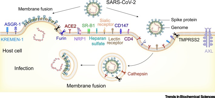
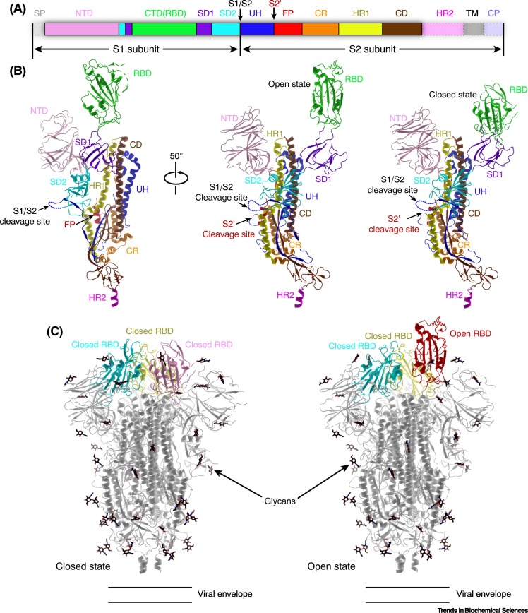
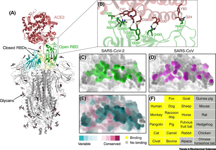
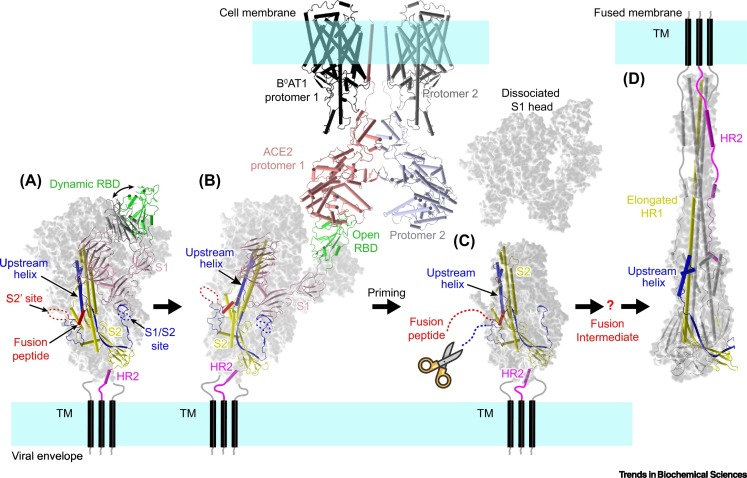
Severe acute respiratory syndrome virus 2 (SARS-CoV-2) invades host cells by interacting with receptors/coreceptors, as well as with other cofactors, via its spike (S) protein that further mediates fusion between viral and cellular membranes. The host membrane protein, angiotensin-converting enzyme 2 (ACE2), is the major receptor for SARS-CoV-2 and is a crucial determinant for cross-species transmission. In addition, some auxiliary receptors and cofactors are also involved that expand the host/tissue tropism of SARS-CoV-2. After receptor engagement, specific proteases are required that cleave the S protein and trigger its fusogenic activity. Here we discuss the recent advances in understanding the molecular events during SARS-CoV-2 entry which will contribute to developing vaccines and therapeutics.

Keywords: COVID-19; SARS-CoV-2; coreceptor; membrane fusion; receptor recognition; spike protein; virus entry.


Conflict of interest statement

Declaration of interests We declare no competing interests.

Figures

Figure I
Figure I
Timeline for the identification of human-infecting coronaviruses (HCoVs).
Figure 1
Figure 1
Schematic diagram of SARS-CoV-2 entry pathways. Multiple molecules at the cell surface are involved in the entry of SARS-CoV-2, including the major receptor ACE2 [2,19], the membrane protease TMPRSS2, and other potential alternative/auxiliary receptors or cofactors [25,52,53,62,70,71,75,80]. Membrane fusion can take place either at the cell surface (left) or in the endosome (right). Both entry pathways are utilized by SARS-CoV-2 [45,46]. Abbreviations: ACE2, angiotensin-converting enzyme 2; SARS-CoV-2, severe acute respiratory syndrome virus 2; TMPRSS2, transmembrane serine protease 2.
Figure 2
Figure 2
Structure of SARS-CoV-2 spike (S) protein. (A) Schematic diagram of the domain organization of S protein. Each domain is represented by a unique color. The signal peptide (SP) and the unresolved region at the C terminus are transparent with dashed outlines. (B) Architecture of an S protomer (PDB: 6VXX and 6VYB). The structure is shown in cartoons and colored by domains as in (A). The unresolved regions are represented by broken lines. (C) Structures of the SARS-CoV-2 S trimer in different conformations. The RBD can adopt different conformations (closed or open) in the S trimer, and only the open conformation is competent for binding to the receptor ACE2 [17,18,26]. Abbreviations: ACE2, angiotensin-converting enzyme 2; CD, central domain; CP, cytoplasmic region; CR, connecting region; CTD, C-terminal domain; FP, fusion peptide; HR1, heptad repeat 1; HR2, heptad repeat 2; NTD, N-terminal domain; RBD, receptor-binding domain; SARS-CoV-2, severe acute respiratory syndrome virus 2; S1/S2 and S2′, cleavage sites within S protein; SD1, subdomain 1; SD2, subdomain 2; TM, transmembrane region; UH, upstream helix.
Figure 3
Figure 3
Interactions between SARS-CoV-2 spike (S) protein and ACE2. (A) Overall structure of a trimeric S bound to ACE2 (PDB: 7KNB). ACE2 binds to the RBD of S in the open conformation, while the other two closed RBDs are inaccessible by the receptor. (B) Close-up view of the S–ACE2 contacting interface (PDB: 6LZG). The key interacting residues are shown as sticks. Hydrogen bonds and salt bridges are represented by dashed lines. (C,D) The S-binding footprint on ACE2 for SARS-CoV-2 (C; PDB: 6LZG) and SARS-CoV (D; PDB: 2AJF). The interaction between SARS-CoV-2 (green) and ACE2 involves more atomic contacts than for SARS-CoV (magenta) [19., 20., 21.,28]. (E) Conservation of the S-binding interface on ACE2. The interface for SARS-CoV-2 S binding displays a high degree of variation among ACE2 orthologs from different animal species. (F) A list of animals tested whose ACE2 can (yellow) or cannot (grey) bind to the RBD of SARS-CoV-2 S protein [29,31,32]. Abbreviations: ACE2, angiotensin-converting enzyme 2; RBD, receptor-binding domain; SARS-CoV-2, severe acute respiratory syndrome virus 2.
Figure 4
Figure 4
Model of the SARS-CoV-2 entry process. (A) The trimeric spike on viral envelope can adopt different conformations (PDB: 6VXX and 6VYB). The RBD undergoes dynamic transition between the closed and open conformations. The SARS-CoV-2 S protein is prone to be cleaved into the S1 and S2 subunits during biosynthesis or cell entry process, and the two subunits remain noncovalently associated to form trimeric spikes [16,17,45]. (B) Only the RBD in the open conformation can bind to the receptor ACE2 (PDB: 7KNB and 6M17). Binding of ACE2 can promote the transition of adjacent RBDs to the open conformation, and thus may facilitate the binding of more ACE2 molecules [33., 34., 35.]. (C) ACE2 binding induces conformational changes that destabilize the interactions between S1 and S2 subunits, thus probably triggering the dissociation of the S1 head [33]. The second proteolysis event at the S2′ site, by TMPRSS2 and other proteases [25,52], produces a free N terminus for the FP and triggers conformational changes to expose the FP for cellular membrane targeting. The question mark indicates the fusion intermediate structures that are not well understood. It is thought that the central domain will refold to form an elongated helix stalk with the HR1 helix, which facilitates FP reaching the target membrane [26]. (D) The HR2 helices then fold upwards to contact the elongated HR1 stalk helices, transforming into the six-helix bundle postfusion conformation [24,26]. This rearrangement draws the viral and cellular membranes into close proximity and induces fusion. Abbreviations: ACE2, angiotensin-converting enzyme 2; B0AT1, sodium-dependent amino acid transporter 1, also known as SLC6A19; FP, fusion peptide; HR1, heptad repeat 1; HR2, heptad repeat 2; RBD, receptor-binding domain; SARS-CoV-2, severe acute respiratory syndrome virus 2; TM, transmembrane region.

Similar articles

Cited by

References

    1. Coronaviridae Study Group of the International Committee on Taxonomy of Viruses The species severe acute respiratory syndrome-related coronavirus: classifying 2019-nCoV and naming it SARS-CoV-2. Nat. Microbiol. 2020;5:536–544. - PMC - PubMed
    1. Zhou P., et al. A pneumonia outbreak associated with a new coronavirus of probable bat origin. Nature. 2020;579:270–273. - PMC - PubMed
    1. Zhu N., et al. A novel coronavirus from patients with pneumonia in China, 2019. New Engl. J. Med. 2020;382:727–733. - PMC - PubMed
    1. Wu F., et al. A new coronavirus associated with human respiratory disease in China. Nature. 2020;579:265–269. - PMC - PubMed
    1. Zhong N.S., et al. Epidemiology and cause of severe acute respiratory syndrome (SARS) in Guangdong, People's Republic of China, in February, 2003. Lancet. 2003;362:1353–1358. - PMC - PubMed

Publication types

Substances