. 2021 Jul 6;118(27):e2106754118.
doi: 10.1073/pnas.2106754118.

B cell residency but not T cell-independent IgA switching in the gut requires innate lymphoid cells

Affiliations

B cell residency but not T cell-independent IgA switching in the gut requires innate lymphoid cells

Mingzhu Zheng et al. Proc Natl Acad Sci U S A. .

Abstract

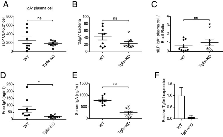
Immunoglobulin A (IgA)-producing plasma cells derived from conventional B cells in the gut play an important role in maintaining the homeostasis of gut flora. Both T cell-dependent and T cell-independent IgA class switching occurs in the lymphoid structures in the gut, whose formation depends on lymphoid tissue inducers (LTis), a subset of innate lymphoid cells (ILCs). However, our knowledge on the functions of non-LTi helper-like ILCs, the innate counter parts of CD4 T helper cells, in promoting IgA production is still limited. By cell adoptive transfer and utilizing a unique mouse strain, we demonstrated that the generation of IgA-producing plasma cells from B cells in the gut occurred efficiently in the absence of both T cells and helper-like ILCs and without engaging TGF-β signaling. Nevertheless, B cell recruitment and/or retention in the gut required functional NKp46-CCR6+ LTis. Therefore, while CCR6+ LTis contribute to the accumulation of B cells in the gut through inducing lymphoid structure formation, helper-like ILCs are not essential for the T cell-independent generation of IgA-producing plasma cells.

Keywords: B cell; IgA; T cell; innate lymphoid cell; lymphoid tissue inducer.


Conflict of interest statement

The authors declare no competing interest.

Figures

Fig. 1.
Fig. 1.
T cell–dependent, but not independent, pathway for IgA class switching requires ROR-γt and ILCs. (A) Flow cytometry analysis and quantification of IgA+ bacteria in WT, Tcrb−/−d−/−, Rorcgfp/gfp, and Rag2−/−Il2rg−/− mice. (B and C) Feces free IgA (B) and serum IgA (C) from above mice detected by ELISA. (D) Flow cytometry analysis and quantification of IgA+ B220 plasma cells in siLP from above mice. (E) The generation of IgA+ B220 plasma cells in siLP from B cells after adoptive transfer were determined by flow cytometry. (F) IgA+ bacteria in E were calculated. (G and H) Feces free IgA (G) and serum IgA (H) from above mice (E) detected by ELISA. Data are representative of at least two independent experiments (AD) or a combination of three independent experiments (EH). Mean ± SD; n = 3 to 10; ns, not significant, *P < 0.05, **P < 0.01, ***P < 0.001, ****P < 0.0001, Student’s t test.
Fig. 2.
Fig. 2.
Functional NKp46CCR6+ LTi/LTi-like cells promote B cell residence in the small intestine. (A) Total CD19+ B cell numbers in the spleen of WT (Gata3fl/fl) and Gata3-deficient (Gata3fl/flVavCre) mice were compared. (B) Total CD19+ B cell numbers in the siLP of these mice were compared. (C and D) CD45.1+ B cells in the spleen (C) and siLP (D) were analyzed and calculated 1 wk after B cell transfer. (E) Imaging of villi in the small intestine of WT, Gata3fl/flVavCre, and Rorcgfp/gfp mice showing the distribution of CD3+ T cells, CD19+ B, and ROR-γt+ ILC3s. (Scale bars, 50 μm.) (F) Flow cytometry analysis of CCR6+ LTi cells from the siLP of WT and Gata3fl/flVavCre mice (Upper) and the quantification of total cell numbers (Lower). (G) Imaging of villi in the small intestine of Gata3fl/fl and Gata3fl/flRorcCre mice showing the distribution of CD3+ T cells, CD19+ B, and ROR-γt + ILC3s. (Scale bars, 50 μm.) (H) The total numbers of CD19+ B cells in the siLP of WT, Gata3fl/flVavCre, and Gata3fl/flVavCre-TghCD2-Gata3 mice were compared. (I) Volcano plot of the differentially expressed genes (fold change >2; P < 0.005) through RNA-Seq analysis of WT and GATA3-deficient LTi cells harvested from the siLP of WT and Gata3fl/flVavCre mice, respectively (39). The positions of some representative genes, including Ret, Il22, Cd4, Il7r, Lta, Ltb, and Ccr6, were labeled in the plot. A full list of differentially expressed genes can be found in Dataset S1. Data are representative of two independent experiments (CG) or three independent experiments (A, B, and H). Mean ± SD; n = 3 to 7; *P < 0.05, **P < 0.01, ***P < 0.001, Student’s t test. Biological duplicates were used for each genotype in the RNA-Seq analysis (I).
Fig. 3.
Fig. 3.
Efficient T cell–independent IgA class switching in the small intestine of Gata3fl/flVavCre mice. (A) Feces free IgAs from WT, Gata3fl/flVavCre, and Rag2−/−Il2rg−/− mice were detected by ELISA. (B) Flow cytometry analysis of IgA+ bacteria in WT, Gata3fl/flVavCre, and Rag2−/−Il2rg−/− mice. (C) Quantification of IgA+ bacteria from mice in B. (D) Serum IgA from above mice detected by ELISA. (E) Flow cytometry analysis of IgA+ B220 plasma cells in siLP from WT and Gata3fl/flVavCre mice. (F) Flow cytometry analysis of CD138 and Blimp1 expression by B220IgA+ plasma cells and B220+IgA cells in E. (G) Quantification of IgA+ B220 plasma cells in E. (H) Quantification of IgA+ B220 plasma cells versus B cell ratio in the siLP. Data are representative of at least two independent experiments (AH). Mean ± SD; n = 3 to 8; ns, not significant, **P < 0.01, ***P < 0.001, ****P < 0.0001, Student’s t test.
Fig. 4.
Fig. 4.
T cell– and ILC-independent IgA switching occurs in the absence of TGF-β signaling. (A) The generation of IgA+ B220 plasma cells in siLP were assessed by flow cytometry in the Rag2−/−Il2rg−/− mice reconstituted with WT or Tgfbr-KO B cells. (B) IgA+ bacteria in A were calculated. (C) Quantification of IgA+ B220 plasma cells to B cell ratio in the siLP. (D and E) Feces free IgAs (D) and serum IgAs (E) from above mice (A) were detected by ELISA. (F) qRT-PCR was carried out to determine the efficiency of Tgfbr1 deletion in Tgfbr-KO splenic B cells by tamoxifen treatment before cell transfer. Data represent the combination of two independent experiments. (AF) Mean ± SD; n = 9; ns, not significant, *P < 0.05, ***P < 0.001, Student’s t test.

Similar articles

Cited by

References

    1. Mowat A. M., Agace W. W., Regional specialization within the intestinal immune system. Nat. Rev. Immunol. 14, 667–685 (2014). - PubMed
    1. Brown E. M., Sadarangani M., Finlay B. B., The role of the immune system in governing host-microbe interactions in the intestine. Nat. Immunol. 14, 660–667 (2013). - PubMed
    1. Buettner M., Lochner M., Development and function of secondary and tertiary lymphoid organs in the small intestine and the colon. Front. Immunol. 7, 342 (2016). - PMC - PubMed
    1. Pearson C., Uhlig H. H., Powrie F., Lymphoid microenvironments and innate lymphoid cells in the gut. Trends Immunol. 33, 289–296 (2012). - PubMed
    1. Caballero-Franco C., et al. ., Epithelial control of gut-associated lymphoid tissue Formation through p38α-dependent restraint of NF-κB signaling. J. Immunol. 196, 2368–2376 (2016). - PMC - PubMed

Publication types

MeSH terms