. 2021 Jun 29;12(1):4024.
doi: 10.1038/s41467-021-24337-9.

The bacterial toxin ExoU requires a host trafficking chaperone for transportation and to induce necrosis

Affiliations

The bacterial toxin ExoU requires a host trafficking chaperone for transportation and to induce necrosis

Vincent Deruelle et al. Nat Commun. .

Abstract

Pseudomonas aeruginosa can cause nosocomial infections, especially in ventilated or cystic fibrosis patients. Highly pathogenic isolates express the phospholipase ExoU, an effector of the type III secretion system that acts on plasma membrane lipids, causing membrane rupture and host cell necrosis. Here, we use a genome-wide screen to discover that ExoU requires DNAJC5, a host chaperone, for its necrotic activity. DNAJC5 is known to participate in an unconventional secretory pathway for misfolded proteins involving anterograde vesicular trafficking. We show that DNAJC5-deficient human cells, or Drosophila flies knocked-down for the DNAJC5 orthologue, are largely resistant to ExoU-dependent virulence. ExoU colocalizes with DNAJC5-positive vesicles in the host cytoplasm. DNAJC5 mutations preventing vesicle trafficking (previously identified in adult neuronal ceroid lipofuscinosis, a human congenital disease) inhibit ExoU-dependent cell lysis. Our results suggest that, once injected into the host cytoplasm, ExoU docks to DNAJC5-positive secretory vesicles to reach the plasma membrane, where it can exert its phospholipase activity.


Conflict of interest statement

The authors declare no competing interests.

Figures
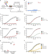
Fig. 1
Fig. 1. DNAJC5 is required for ExoU cytotoxicity.
a Screening process to identify host genes required for ExoU toxicity. A gRNA library (TKOv3, four gRNAs per gene) was constructed for A549 human epithelial cells. Cells were subjected to three 90-min rounds of infection with the ExoU+ PA14 strain in triplicates. Infection was stopped by washing and adding antibiotics. DNA, corresponding to gRNAs from resistant cells and from the uninfected library, were then amplified by PCR and submitted to deep sequencing. b Analysis of sequencing data. Statistical analysis of gRNA abundance in infected vs uninfected conditions. gRNAs targeting the DNAJC5 gene were the only ones significantly enriched in the screen. c Cytotoxicity assay. A clonal population of DNAJC5−/− A549 cells, native A549 cells or cells transfected with an empty vector (EV) were infected in triplicates with PA14 and necrosis was monitored by propidium iodide incorporation, recorded by time-lapse microscopy. Results are represented as the mean percentage (±SD) of necrotic cells. d Cytotoxicity assay with A549 native cells. DNAJC5−/− cells complemented with DNAJC5 (DNAJC5−/−::DNAJC5), or the mock-complemented control, were infected with PA14. Results (n = 4 replicates) are represented as the mean percentage (±SD) of necrotic cells. e, f Cytotoxicity assays and representation are similar to (c,d), but cells were infected with the ExoU+ PP34 strain (n = 3 and 4 for (e) and (f), respectively). g T3SS-dependent injection of ExoU in A549 epithelial cells. A549 or DNAJC5−/− cells were infected with PP34ΔexoU expressing ExoUS142A fused to the β-lactamase (Bla) or infected with uncomplemented PP34ΔexoU. Cells were loaded with CCF2, a fluorescent substrate for Bla, which shifts from green to blue fluorescence upon processing by the enzyme. Fluorescence was measured at 4 hpi on both channels and results are expressed as a blue/green ratio (n = 3; bar: median). Statistical differences between data in A549 and DNAJC5−/− cells infected with the exoU-bla strain were calculated with a two-sided Student’s test, and were not significant (n.s.). h Cytotoxicity assay. Increasing concentrations of quercetin were added to A549 cells in the presence of PA14 (ExoU+) at an MOI of 10. Necrosis was monitored by PI incorporation and recorded by time-lapse microscopy. Results (n = 8 replicates) are represented as the mean percentage (±SD) of necrotic cells. Source data are provided as a Source Data file.
Fig. 2
Fig. 2. ExoU toxicity in Drosophila requires CSP (DNAJC5 orthologue).
a Drosophila were infected with PP34 (ExoU+) by pricking the thorax with a thin needle dipped in bacterial suspension. b Flies expressing RNAi transgenes targeting either the firefly luciferase gene (Luc-KD, n = 33) or the Csp gene (CSP-KD1, n = 40) were infected with PP34. c Similar experiment with Luc-KD (n = 28) and CSP-KD2 (n = 51) infected with PP34. Mock-infected CSP-KD1 (b, n = 30) or CSP-KD2 (c, n = 47) flies were included as negative control (uninfected). Fly survival was recorded and data are represented as Kaplan–Meyer curves. Multiple comparisons tests (LogRank) gave a p-value of 0.0001 for (b, c). Simple LogRank comparison tests were performed: n.s., non-significant; ***p < 0.0001. Source data are provided as a Source Data file.
Fig. 3
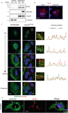
Fig. 3. ExoU localizes in DNAJC5-positive vesicles.
a Fractionation of DNAJC5−/− (KO) and DNAJC5−/−::DNAJC5 (WT) cells infected with a P. aeruginosa’s strain (CHA-exoUS142A) secreting a catalytically inactive ExoU mutant through its T3SS. Cells were harvested at 4 hpi and their soluble and membrane fractions were prepared. Western blots were performed on fractions using anti-ExoU, DNAJC5, Lamp2 (a late endosome-lysosome marker) and β-tubulin (a cytosolic marker) antibodies. The higher molecular weight bands revealed by the ExoU antibody represent the ubiquitinylated form of ExoU. Source data are provided as a Source Data file. b ExoU immunofluorescence staining of A549 and DNAJC5−/− cells infected with CHA-exoUS142A. A single representative z-section is shown. c left DNAJC5-FLAG immunofluorescence signals (green) in uninfected DNAJC5−/−::DNAJC5 cells. Four z-sections obtained by confocal microscopy are shown from top to bottom. A z-projection is also shown below. Nuclei were counterstained in blue. c right DNAJC5-FLAG (green) and ExoU (red) immunofluorescence signals in DNAJC5−/−::DNAJC5 cells infected with CHA-exoUS142A. As for uninfected cells, four z-sections and a z-projection are shown. For each section, a region was enlarged and an arrow was drawn (represented on the right) to establish an intensity profile for both green and red fluorescences, as shown. d DNAJC5-GFP and ExoU localizations in transfected HUVEC infected with CHA-exoUS142A on a wide-field microscopy image. Arrowheads show colocalization of both markers. The insert is an enlargement of the merged image, showing DNAJC5 and ExoU localization at the vesicle’s membrane.
Fig. 4
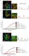
Fig. 4. DNAJC5 co-chaperone activity is dispensable for ExoU toxicity.
a Domain organization of the human DNAJC5 protein. The following locations are highlighted: the two phosphorylation sites (serine 10 and serine 34), the HPD motif for Hsc70/Hsp70 binding, and the cysteine-rich region containing the leucines L115 and L116, mutated in adult neuronal ceroid lipofuscinosis patients (stars). b Effect of DNAJC5 mutations, H43Q and S10A-S34A, on ExoU toxicity. A549 cells, as well as DNAJC5−/− cells complemented with either DNAJC5, DNAJC5H43Q, DNAJC5S10A-S34A or the empty vector (EV) were subjected to an infection assay with PA14. Data are shown as the mean ± SD. N = 6 fields per condition. c DNAJC5-FLAG (green) and ExoU (red) immunofluorescence signals for DNAJC5−/−::DNAJC5H43Q and DNAJC5−/−::DNAJC5S10A-S34A cells infected with CHA-exoUS142A strain, which secretes a catalytically inactive ExoU mutant. One z-section is shown. For each section, a region was enlarged and an arrow was drawn (represented on the right) to establish the intensity profiles for both green and red fluorescences. d, e. Effect of decreased Hsc70 and Hsp70 expression on ExoU toxicity. A549 cells were transfected with siRNAs for Hsp70 (si_Hsp70) or Hsc70 (si_Hsc70) to knock down their expression. Knockdown was monitored by Western blot (d). KD cells were subjected to an infection assay (e)(n = 5), in the same conditions as in (b). Source data are provided as a Source Data file.
Fig. 5
Fig. 5. Trafficking of DNAJC5-positive vesicles is required for ExoU toxicity.
a Localizations by immunofluorescence of ExoU (red) and DNAJC5-FLAG (green) in DNAJC5−/−::DNAJC5L115R cells infected with CHA-exoUS142A on a z-section. A selected area was enlarged and an arrow was drawn, to establish the intensity profiles for both signals. b Immunofluorescence localizations of ExoU (red) and DNAJC5-FLAG (green) in DNAJC5−/−::DNAJC5ΔL116 cells infected with CHA-exoUS142A. Intensity profiles were obtained as above. c Cytotoxicity assay with DNAJC5−/−::DNAJC5L115R and DNAJC5−/−::DNAJC5ΔL116 cells, alongside controls, infected with PA14. Data are shown as the mean ± SD. N = 7 fields per condition. d Immunofluorescence localizations of ExoU (red) and DNAJC5 (green) in DNAJC5−/−::DNAJC5ΔJdomain cells infected with CHA-exoUS142A. Intensity profiles were obtained as above. e Effect of J domain deletion. Control cells and DNAJC5−/− cells complemented with full-length DNAJC5, DNAJC5ΔJdomain or EV were infected with PA14, and cytotoxicity was recorded (mean ± SD; n = 5). Source data are provided as a Source Data file.
Fig. 6
Fig. 6. ExoU phospholipase activity is independent of DNAJC5.
Phospholipase activity of purified ExoU (65 pmols) was measured in the presence of soluble (Sol) or membrane (Mb) fractions from uninfected DNAJC5−/− (KO) or DNAJC5−/−::DNAJC5 (WT) cells. Experiments were performed in triplicates and incubated for 24 h. Data are expressed in nmoles of substrate hydrolyzed per nmoles of ExoU (n = 3; bar: median). Statistical differences were established by two-sided ANOVA (p < 0.0001), followed by Tukey’s test (*p < 0.0001, n.s., non-significant). Source data are provided as a Source Data file.
Fig. 7
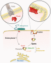
Fig. 7. Proposed model of ExoU trafficking in host cells.
Upon delivery into the host cytoplasm by the T3SS, the toxin uses an endocytic pathway to reach the perinuclear region, as suggested by ExoU/EEA1 colabeling. Then, ExoU binds to the late endosome’s limiting membrane (decorated by Lamp2 and DNAJC5), thanks to the interaction of ExoU with an as yet unidentified specific receptor at the vesicle’s surface. ExoU remains at the external side of the vesicle’s membrane and co-opts the DNAJC5-dependent MAPS pathway to achieve anterograde transport toward the cellular periphery (where vesicles loose Lamp2), and eventually the plasma membrane (PM). Fusion of vesicles with the PM brings ExoU close to PM’s inner leaflet, where its membrane localization domain binds to PI(4,5)P2. PI(4,5)P2 binding triggers conformational changes in ExoU, leading to toxin oligomerization and activation of its phospholipase activity, which eventually induces PM rupture.

Similar articles

Cited by

References

    1. Gellatly SL, Hancock RE. Pseudomonas aeruginosa: new insights into pathogenesis and host defenses. Pathog. Dis. 2013;67:159–173. doi: 10.1111/2049-632X.12033. - DOI - PubMed
    1. Hauser AR. The type III secretion system of Pseudomonas aeruginosa: infection by injection. Nat. Rev. Microbiol. 2009;7:654–665. doi: 10.1038/nrmicro2199. - DOI - PMC - PubMed
    1. Williams BJ, Dehnbostel J, Blackwell TS. Pseudomonas aeruginosa: host defence in lung diseases. Respirology. 2010;15:1037–1056. doi: 10.1111/j.1440-1843.2010.01819.x. - DOI - PubMed
    1. Botelho J, Grosso F, Peixe L. Antibiotic resistance in Pseudomonas aeruginosa—mechanisms, epidemiology and evolution. Drug Resist Updat. 2019;44:100640. doi: 10.1016/j.drup.2019.07.002. - DOI - PubMed
    1. Horcajada, J. P. et al. Epidemiology and treatment of multidrug-resistant and extensively drug-resistant Pseudomonas aeruginosa infections. Clin. Microbiol. Rev.32, 10.1128/CMR.00031-19 (2019). - PMC - PubMed

Publication types

MeSH terms

Substances