. 2021 Jun 29;12(1):4028.
doi: 10.1038/s41467-021-24282-7.

Crystal structure of an archaeal CorB magnesium transporter

Affiliations

Crystal structure of an archaeal CorB magnesium transporter

Yu Seby Chen et al. Nat Commun. .

Abstract

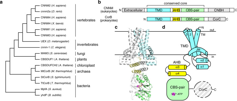
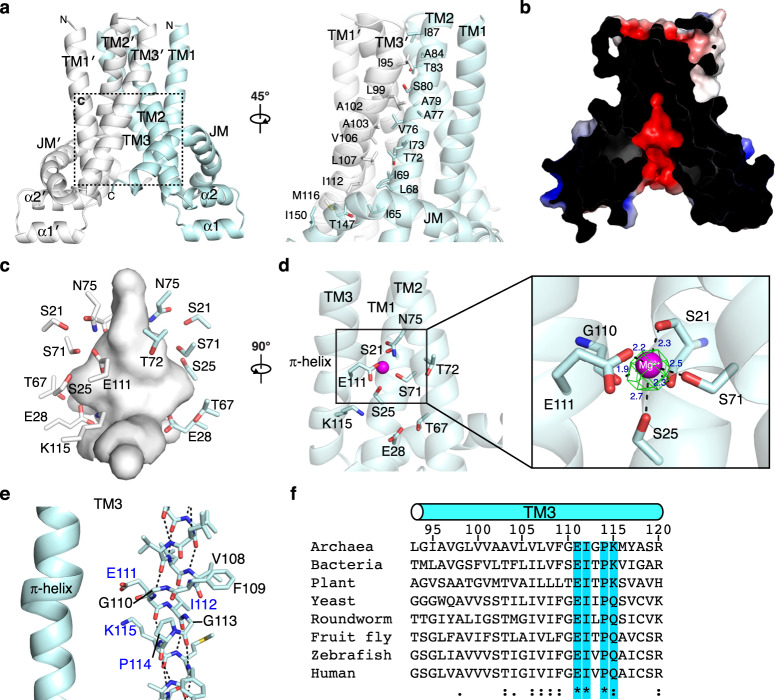
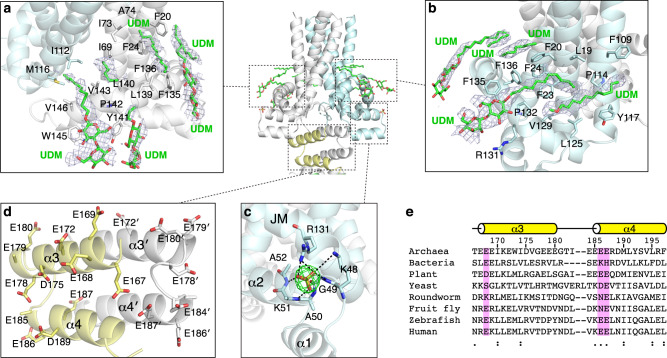
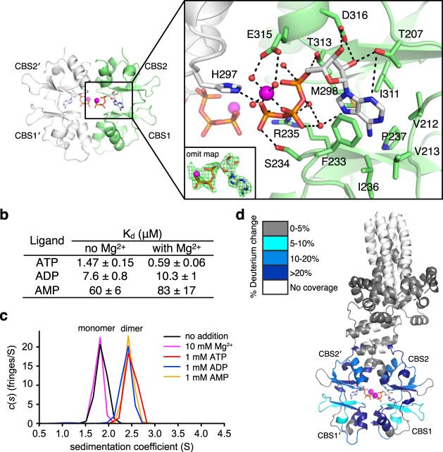
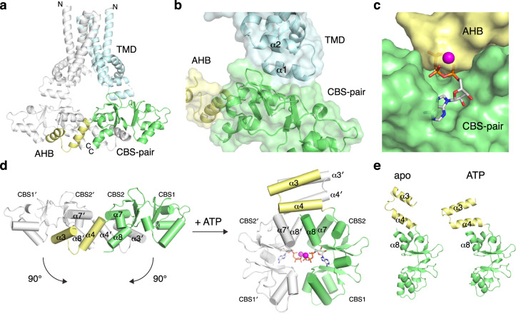
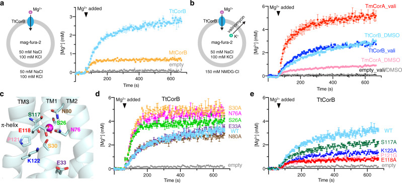
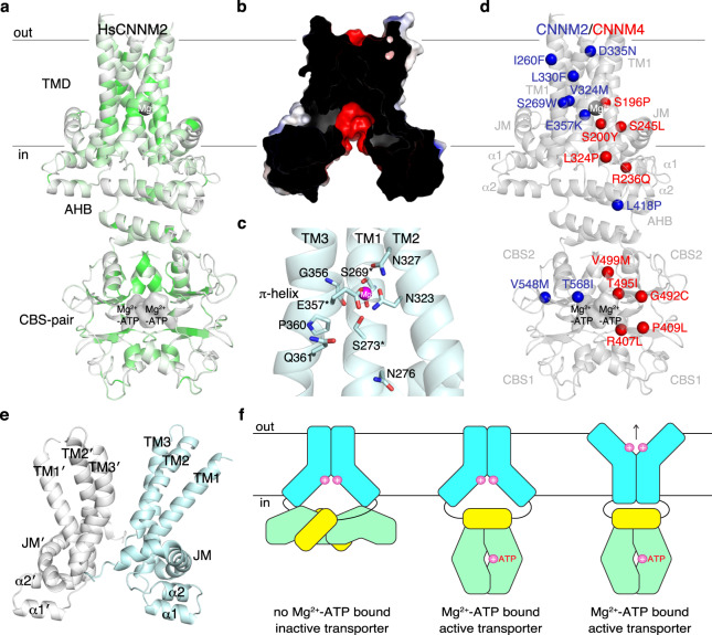
CNNM/CorB proteins are a broadly conserved family of integral membrane proteins with close to 90,000 protein sequences known. They are associated with Mg2+ transport but it is not known if they mediate transport themselves or regulate other transporters. Here, we determine the crystal structure of an archaeal CorB protein in two conformations (apo and Mg2+-ATP bound). The transmembrane DUF21 domain exists in an inward-facing conformation with a Mg2+ ion coordinated by a conserved π-helix. In the absence of Mg2+-ATP, the CBS-pair domain adopts an elongated dimeric configuration with previously unobserved domain-domain contacts. Hydrogen-deuterium exchange mass spectrometry, analytical ultracentrifugation, and molecular dynamics experiments support a role of the structural rearrangements in mediating Mg2+-ATP sensing. Lastly, we use an in vitro, liposome-based assay to demonstrate direct Mg2+ transport by CorB proteins. These structural and functional insights provide a framework for understanding function of CNNMs in Mg2+ transport and associated diseases.


Conflict of interest statement

The authors declare no competing interests.

Figures

Fig. 1
Fig. 1. Overall structure and domain organization of CNNM/CorB Mg2+ transporters.
a Phylogenetic analysis of representative CNNM/CorB orthologs generated using the neighbor-joining method. b Domain organization of eukaryotic CNNM and prokaryotic CorB. TMD transmembrane domain, AHB acidic helical bundle, CNBH cyclic nucleotide-binding homology domain, CorC cobalt resistance C domain. c Crystal structure of the Mg2+-ATP-bound MtCorB without the C-terminal CorC domain as a homodimer. One chain is colored by domains. d Topology of a MtCorB monomer showing the transmembrane and juxtamembrane helices of the TMD (residues 1–154, cyan), the two helices of the AHB (residues 166–199, yellow), the Mg2+-ATP-binding CBS-pair domain (residues 200–324, green), and the CorC domain (residues 325–426, grey).
Fig. 2
Fig. 2. TM domain in an inward-facing conformation with a negatively charged cavity.
a TM domain homodimerizes with interface formed by TM2 and TM3 of each protomer. b Electrostatic surface potential representation (±5 kT e−1) of MtCorB TM domain showing a cross-sectional view of the negatively charged cavity. c Close-up view of the polar residues forming the cavity. d A magnesium ion (Mg2+) bound in the cavity with FoFc omits map contoured at 5.0σ. e π-helical turn preceding Pro114 in TM3. Highly conserved residues are highlighted in blue. f Conservation of residues in the π-helical turn from archaea to humans.
Fig. 3
Fig. 3. Juxtamembrane helix and acidic helical bundle.
a, b UDM detergent molecules bound by the juxtamembrane helix. Simulated annealing 2FoFc composite omit map contoured at 1.0σ. c A sulfate ion bound between JM and helix–turn–helix motif with FoFc omit map contoured at 5.0σ. d Conservation of acidic residues in the AHB domain. e Sequence conservation of conserved glutamates in the AHB.
Fig. 4
Fig. 4. Mg2+-ATP binding to CBS-pair domain.
a Structural basis of Mg2+-ATP binding. Mg2+ ions and water molecules are shown in magenta and red, respectively. The Mg2+-ATP FoFc omits map was contoured at 3.0σ. b Affinities of MtCorBΔC to adenosine nucleotides with and without 50 mM Mg2+ measured by ITC. c Dimerization of the MtCorB CBS-pair domain in the presence of 1 mM adenosine nucleotides as measured by SV-AUC experiments. d Conformational change in the MtCorBΔC in the presence and absence of Mg2+-ATP measured by HDX-MS. Regions that showed significant decreases in exchange (defined as >5%, 0.4 Da, and a two-tailed Student’s t test p < 0.01) in the presence of Mg2+-ATP are colored blue. Peptides in the CBS-pair domain dimerization interface show the most protection from deuterium exchange upon Mg2+-ATP binding.
Fig. 5
Fig. 5. Apo conformation of MtCorBΔC captured by R235L mutant.
a Overall structure of MtCorBΔC R235L mutant. One chain is colored by domains. b Novel domain–domain contact between TMD and CBS-pair domain. c AHB binds to CBS-pair domain and competes for Mg2+-ATP site. d Large conformational change in the AHB and CBS-pair domains upon Mg2+-ATP binding. e The apo and ATP-bound structures are similar with differences limited to the AHB and α8 helix.
Fig. 6
Fig. 6. Liposome-based assay shows direct Mg2+ transport by CorB proteins.
a Liposomal assay showing direct transport of Mg2+ by MtCorB and TtCorB. Proteoliposomes (protein/lipid ratio of 1:30) or empty liposomes containing mag-fura-2 were equilibrated for 1 min before the addition of 5 mM MgCl2 to initiate Mg2+ uptake (arrowhead). The data points represent mean ± SEM (n = 3 independent measurements). b An inward negative membrane potential generated by valinomycin leads to enhanced Mg2+ transport by TmCorA (protein/lipid ratio of 1:270) but not TtCorB (protein/lipid ratio of 1:30), suggesting electroneutral transport of Mg2+ by TtCorB. The data points represent mean ± SEM (n = 3 independent measurements). c Homology model of TtCorB showing locations of conserved residues in the negatively charged cavity and Mg2+ binding site. d, e Liposomal Mg2+ transport assays of TtCorB mutants under the same experimental conditions as panel (a). Mutations in the Mg2+-binding site increased transport activity while mutations near the π-helical turn reduced transport. The data points represent mean ± SEM (n = 3 independent measurements).
Fig. 7
Fig. 7. Molecular modeling.
a Homology model of human CNNM2 (HsCNNM2) color-coded according to identity to CNNM/CorB proteins shown in Supplementary Fig. 2. Conservation is highest around the Mg2+ binding site, negatively charged cavity, and the CBS-pair dimerization interface. b Electrostatic surface potential analysis (±5 kT e−1) shows conservation of negatively charged cavity in HsCNNM2 in cross-sectional view. c Conservation of the polar residues in the Mg2+ binding site and π-helical turn of HsCNNM2. Asterisk denotes disease-causing mutations. d Mutations in CNNM2 and CNNM4 responsible for hypomagnesemia and Jalili syndrome cluster around the core of the TMD and nucleotide-binding site. For clarity, mutations are only shown on one chain. e Outward-facing conformation generated by MD simulations. f Proposed model of MtCorB transport and regulation. Loss of Mg2+-ATP binding, CBS-TMD contact inactivates TMD in the inward-facing conformation. Upon Mg2+-ATP binding, CBS-TMD contact is alleviated, thereby activating and allowing TMD to mediate Mg2+ transport.

References

    1. de Baaij JH, Hoenderop JG, Bindels RJ. Magnesium in man: implications for health and disease. Physiol. Rev. 2015;95:1–46. doi: 10.1152/physrev.00012.2014. - DOI - PubMed
    1. Funato Y, Miki H. Molecular function and biological importance of CNNM family Mg2+ transporters. J. Biochem. 2019;165:219–225. doi: 10.1093/jb/mvy095. - DOI - PubMed
    1. Stuiver M, et al. CNNM2, encoding a basolateral protein required for renal Mg2+ handling, is mutated in dominant hypomagnesemia. Am. J. Hum. Genet. 2011;88:333–343. doi: 10.1016/j.ajhg.2011.02.005. - DOI - PMC - PubMed
    1. Yamazaki D, et al. Basolateral Mg2+ extrusion via CNNM4 mediates transcellular Mg2+ transport across epithelia: a mouse model. PLoS Genet. 2013;9:e1003983. doi: 10.1371/journal.pgen.1003983. - DOI - PMC - PubMed
    1. Arjona FJ, et al. CNNM2 mutations cause impaired brain development and seizures in patients with hypomagnesemia. PLoS Genet. 2014;10:e1004267. doi: 10.1371/journal.pgen.1004267. - DOI - PMC - PubMed

Publication types

MeSH terms

Supplementary concepts