. 2021 Jun 29;11(1):13451.
doi: 10.1038/s41598-021-92812-w.

Noncytotoxic silver nanoparticles as a new antimicrobial strategy

Affiliations

Noncytotoxic silver nanoparticles as a new antimicrobial strategy

Bartosz Skóra et al. Sci Rep. .

Abstract

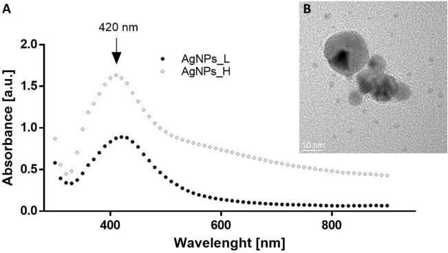
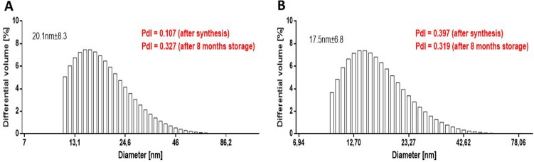
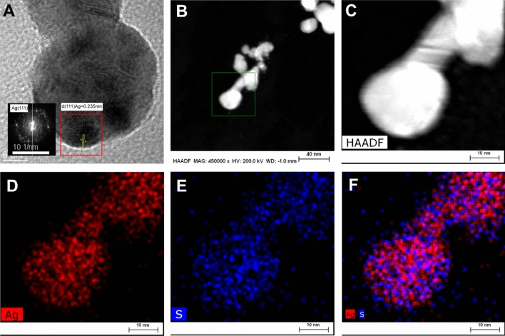
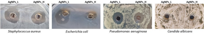
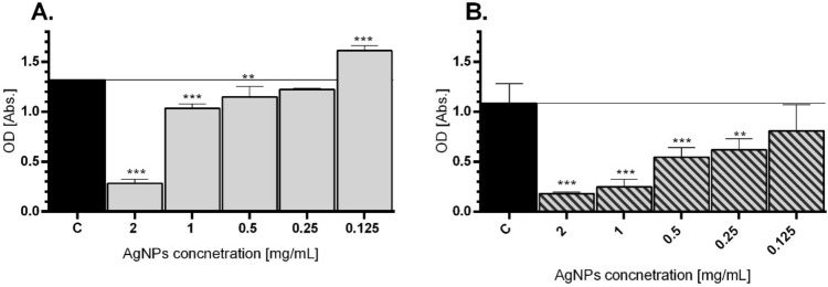
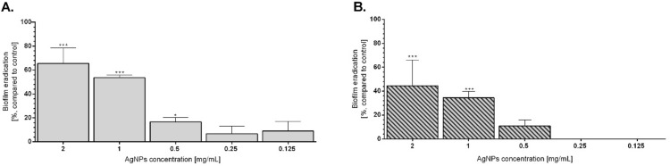
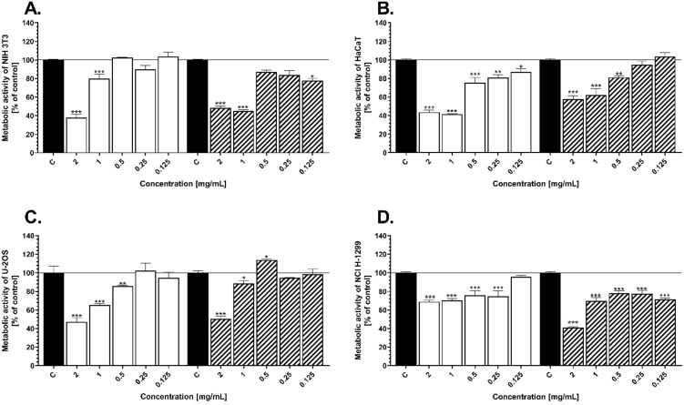
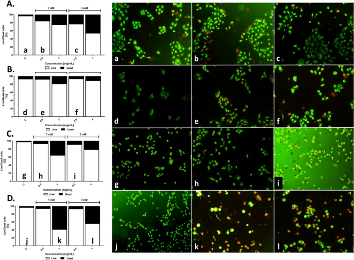
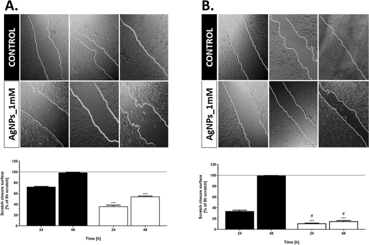
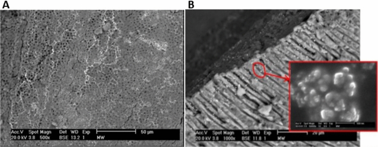
Drug-resistance of bacteria is an ongoing problem in hospital treatment. The main mechanism of bacterial virulency in human infections is based on their adhesion ability and biofilm formation. Many approaches have been invented to overcome this problem, i.e. treatment with antibacterial biomolecules, which have some limitations e.g. enzymatic degradation and short shelf stability. Silver nanoparticles (AgNPs) may be alternative to these strategies due to their unique and high antibacterial properties. Herein, we report on yeast Saccharomyces cerevisiae extracellular-based synthesis of AgNPs. Transmission electron microscopy (TEM) revealed the morphology and structure of the metallic nanoparticles, which showed a uniform distribution and good colloid stability, measured by hydrodynamic light scattering (DLS). The energy dispersive X-ray spectroscopy (EDS) of NPs confirms the presence of silver and showed that sulfur-rich compounds act as a capping agent being adsorbed on the surface of AgNPs. Antimicrobial tests showed that AgNPs inhibit the bacteria growth, while have no impact on fungi growth. Moreover, tested NPs was characterized by high inhibitory potential of bacteria biofilm formation but also eradication of established biofilms. The cytotoxic effect of the NPs on four mammalian normal and cancer cell lines was tested through the metabolic activity, cell viability and wound-healing assays. Last, but not least, ability to deep penetration of the silver colloid to the root canal was imaged by scanning electron microscopy (SEM) to show its potential as the material for root-end filling.


Conflict of interest statement

The authors declare no competing interests.

Figures

Figure 1
Figure 1
(A) UV–Vis absorption spectra of biosynthesized AgNPs_L (circle) and AgNPs_H (diamond). (B) Example of TEM image of AgNPs.
Figure 2
Figure 2
Size of the AgNPs_L (A) and AgNPs_H (B) evaluated by hydrodynamic light scattering analysis. PdI, polidyspersity index.
Figure 3
Figure 3
(A) Representative HRTEM images of single silver nanoparticle with lattice fringes. Silver was identified by the inter-planes spacing d = 0.235 nm corresponding to the (111) plane of silver. Measurement of inter-planes spacing was done on FFT image. (B, C) HAADF STEM image of AgNPs with various magnification. (DF) The chemical composition analysis of AgNPs. EDS elemental mapping images of silver (red), sulfur (blue), and an overlay of the Ag and S. All the EDS measurements are collected from image B (lower magn.) and C (higher magn).
Figure 4
Figure 4
Antimicrobial activity of the AgNPs after 24 h of incubation against Staphylococcus aureus, Escherichia coli, Pseudomonas aeruginosa and Candida albicans.
Figure 5
Figure 5
Biofilm inhibition after treatment of AgNPs in E. coli (A) and P. aeruginosa (B). Strains were incubated for 24 h in the presence of AgNPs. Post-treatment surface-associated biofilm was stained and the OD of biofilm biomass were presented. Mean values with standard deviation (error bars) with *, **, ***are statistically different from the respective control at P < 0.05, P < 0.01, and P < 0.001, respectively (one-way ANOVA, Tukey test).
Figure 6
Figure 6
Biofilm eradication after treatment of AgNPs in E. coli (A) and P. aeruginosa (B). Created biofilms were treated with AgNPs for 24 h and the percentage of biofilm eradication in comparison to untreated bacteria was calculated. Mean values with standard deviation (error bars) with *, **, ***are statistically different from the respective control at P < 0.05, P < 0.01, and P < 0.001, respectively (one-way ANOVA, Tukey test).
Figure 7
Figure 7
Cell metabolic activity of the NIH 3T3 (A), HaCaT (B), U-2OS (C), and NCI-1299 (D) after 24 h exposure to various concentration of AgNPs_L and AgNPs_H. The values are the means (n = 6) with standard deviation (error bars). The statistical significance is indicated as follows: *P < 0.05, **P < 0.01, ***P < 0.005 according to one-way ANOVA, Tukey test.
Figure 8
Figure 8
Cell viability of the NIH3T3 (A), HaCaT (B), U2OS (C) and NCI-1299 (D) after exposure to AgNPs_L and AgNPs_H. Cells after NPs treatment were stained with acridine orange and ethidium bromide, and were imaged with inverted fluorescence microscope (representative images—right panel). Dead cells were scored per 100 total cells analyzed and expressed as % (graphs).
Figure 9
Figure 9
Cell migration ability in response to AgNPs. Comparison of migration in both HaCaT (A) and U-2OS (B) cell lines by taking images at different time intervals (0, 24, 48 h). Results are represented by marking the scratch with parallel lines and visually displaying the number of cells migrated in to the scratch area. The widths were measured using Image J software, and the data were analyzed using Prism 5.0. The values are the means with standard deviation (error bars). The statistical significance is indicated as follows: *P < 0.05, **P < 0.01, ***P < 0.005 according to one-way ANOVA, Tukey test (compared to respective control), and #P < 0.05 are statistically different from respectively tested group (HaCaT vs. U-2OS).
Figure 10
Figure 10
The wall of the macrocanal covered with silver nanoparticles (A) and the micrograp of the penetration of silver nanoparticles into the dentinal canal (B) with agglomerates of nanoparticles (inset).

Similar articles

Cited by

References

    1. Zhang XF, Liu ZG, Shen W, Gurunathan S. Silver nanoparticles: synthesis, characterization, properties, applications, and therapeutic approaches. Int. J. Mol. Sci. 2016 doi: 10.3390/ijms17091534. - DOI - PMC - PubMed
    1. Ahamed M, AlSalhi MS, Siddiqui MKJ. Silver nanoparticle applications and human health. Clin. Chim. Acta. 2010 doi: 10.1016/j.cca.2010.08.016. - DOI - PubMed
    1. Ahmad A, Wei Y, Syed F, Tahir K, Rehman AU, Khan A, Ullah S, Yuan Q. The effects of bacteria-nanoparticles interface on the antibacterial activity of green synthesized silver nanoparticles. Microb. Pathog. 2017 doi: 10.1016/j.micpath.2016.11.030. - DOI - PubMed
    1. Niknejad F, Nabili M, Ghazvini RD, Moazeni M. Green synthesis of silver nanoparticles: Another honor for the yeast model Saccharomyces cerevisiae. Curr. Med. Mycol. 2015 doi: 10.18869/acadpub.cmm.1.3.17. - DOI - PMC - PubMed
    1. Verderosa AD, Totsika M, Fairfull-Smith KE. Bacterial biofilm eradication agents: a current review. Front. Chem. 2019 doi: 10.3389/fchem.2019.00824. - DOI - PMC - PubMed

MeSH terms

LinkOut - more resources