. 2021 Jun 29;11(1):13462.
doi: 10.1038/s41598-021-92434-2.

Reporter cell assay for human CD33 validated by specific antibodies and human iPSC-derived microglia

Affiliations

Reporter cell assay for human CD33 validated by specific antibodies and human iPSC-derived microglia

Jannis Wißfeld et al. Sci Rep. .

Abstract

CD33/Sialic acid-binding Ig-like lectin 3 (SIGLEC3) is an innate immune receptor expressed on myeloid cells and mediates inhibitory signaling via tyrosine phosphatases. Variants of CD33 are associated with Alzheimer's disease (AD) suggesting that modulation of CD33 signaling might be beneficial in AD. Hence, there is an urgent need for reliable cellular CD33 reporter systems. Therefore, we generated a CD33 reporter cell line expressing a fusion protein consisting of the extracellular domain of either human full-length CD33 (CD33M) or the AD-protective variant CD33ΔE2 (D2-CD33/CD33m) linked to TYRO protein tyrosine kinase binding protein (TYROBP/DAP12) to investigate possible ligands and antibodies for modulation of CD33 signaling. Application of the CD33-specific antibodies P67.6 and 1c7/1 to the CD33M-DAP12 reporter cells resulted in increased phosphorylation of the kinase SYK, which is downstream of DAP12. CD33M-DAP12 but not CD33ΔE2-DAP12 expressing reporter cells showed increased intracellular calcium levels upon treatment with CD33 antibody P67.6 and partially for 1c7/1. Furthermore, stimulation of human induced pluripotent stem cell-derived microglia with the CD33 antibodies P67.6 or 1c7/1 directly counteracted the triggering receptor expressed on myeloid cells 2 (TREM2)-induced phosphorylation of SYK and decreased the phagocytic uptake of bacterial particles. Thus, the developed reporter system confirmed CD33 pathway activation by CD33 antibody clones P67.6 and 1c7/1. In addition, data showed that phosphorylation of SYK by TREM2 activation and phagocytosis of bacterial particles can be directly antagonized by CD33 signaling.


Conflict of interest statement

MM, PP, AC, LR, LP and OB are employed by a company with activities related to the topic of this manuscript. MM and OB are named inventors of a submitted patent related to upscaled production of iPSC-derived microglia and macrophages (EP20162230) that is assigned to the LIFE & BRAIN GmbH. OB and HN are named inventors on a patent related to generation of microglial precursors from pluripotent stem cells (patent family to WO2010125107A1) that is assigned to the LIFE & BRAIN GmbH and the University of Bonn. JW and OM declare no potential conflict of interest.

Figures

Figure 1
Figure 1
CD33 reporter cell line constructs. (a) Schematic drawing of the two CD33-DAP12 constructs. Both, the full CD33 ecto-domain (CD33M) and the ecto-domain lacking the sialic acid binding domain (CD33ΔE2) were fused to TYROBP/DAP12. (b) Schematic drawing of the readouts for the CD33 reporter cell line. CD33 can be activated by ligands or antibodies, which results in increased SYK phosphorylation and consequently increased intracellular calcium levels. (c) Gel electrophoresis image of CD33-DAP12 constructs cloned into pcDNA5/FRT after digestion by EcoRI. (d) Successful exchange of the viral CMV promoter with the human EEF1A1 promoter was indicated by a second band after digestion with XhoI at 1,339 bp (CD33M) or 958 bp (CD33ΔE2). (e) Gel electrophoresis image of pcDNA5/FRT-CD33-DAP12-GCaMP6m plasmids after digestion with XhoI. GCaMP6m positive clones exhibited three bands compared to the control (Ctrl) with only two bands. Gel images were cropped for better visualization. Supplementary Fig. 1 shows the uncropped full-length gel.
Figure 2
Figure 2
Flow cytometric analysis of CD33 surface expression. (a) Schematic drawing of the CD33-DAP12 constructs. Both, the full CD33 ecto-domain (CD33M) and the ecto-domain lacking the sialic acid binding domain (CD33∆E2) were fused to TYROBP/DAP12. CD33∆E2 can be identified by binding of the antibody clone 1c7/1 (blue) but not WM53 or P67.6 (red), whereas all three antibody clones can bind CD33M. (b) The CD33-DAP12 and CD33-DAP12-GCaMP6m cells were stained for CD33 surface expression with the antibody clones 1c7/1, WM53 and P67.6. A representative flow cytometry histogram plot for the CD33M-DAP12-GCaMP6m cells is shown (left side). All three tested antibodies were able to stain full-length CD33 on the cell surface. Expression of variant 2 CD33 from CD33∆E2-DAP12 and CD33∆E2-DAP12-GCaMP6m cells was only detected by antibody clone 1c7/1. A representative flow cytometry histogram plot for the CD33∆E2-DAP12-GCaMP6m cells is shown (right side). (c) Quantification of CD33 staining showed a high percentage of CD33 expressing cells in the CD33M-DAP12-GCaMP6m line for all three tested antibody clones but only the CD33 antibody clone 1c7/1 was able to detect CD33 in CD33∆E2-DAP12-GCaMP6m expressing cells. The antibody clones WM53 and P67.6 did not show any staining of CD33∆E2-DAP12 expressing cells. (d) Quantification of CD33 staining revealed a high percentage of cells in the CD33M-DAP12 line expressed CD33, and was detected by all three antibody clones. CD33 in CD33ΔE2-DAP12 expressing cells was only detected by antibody clone 1c7/1. Data are shown as mean + SEM of three to five independent experiments; *** p ≤ 0.001 compared to Secondary Control determined by Welch ANOVA followed by Games-Howell post hoc test.
Figure 3
Figure 3
pSYK detection in CD33M-DAP12 cell lines. (a) pSYK detection in CD33M-DAP12 reporter cells treated with CD33-specific antibodies. Addition of CD33 antibodies P67.6 and 1c7/1 resulted in increased pSYK levels, whilst P67.6 F(ab), WM53 as well as the different isotype IgG1 control antibodies did not show any change in endogenous pSYK levels. (b) 1c7/1 and P67.6 dose–response curve in CD33M-DAP12 reporter cells. Addition of CD33 antibody clones 1c7/1 and P67.6 resulted in an increase in endogenous pSYK levels measured 30 min after the treatment. Data are presented as mean ± or + SD; ** p ≤ 0.01 compared to untreated determined by one-way ANOVA analysis followed by Dunnett’s post hoc test.
Figure 4
Figure 4
Calcium imaging in CD33-DAP12-GCaMP6m reporter cell lines. (a) Schematic time line of image acquisition and compound handling. (b, c) Calcium imaging analyzed as ΔF/F(t) in CD33-DAP12-GCaMP6m lines. Addition 100 µM dATP led to a strong increase in intracellular calcium levels in both cell lines, CD33M- and CD33∆E2-DAP12-GCaMP6m, with a peak at around 20–25 s. The CD33 antibody clones 1c7/1 and P67.6 evoked a selective intracellular calcium response only in CD33M-DAP12-GCaMP6m cells. The CD33 antibody clones WM53 and P67.6 F(ab) as well as the isotype IgG1/F(ab’)2 antibodies did not show a change in intracellular calcium levels. (d, e) The area under the curve as well as the maximum ΔF/F(t) signal calculated from independent experiments showed a significant increase in dATP treated samples in both CD33-DAP12-GCaMP6m lines and a selective increase in CD33M-DAP12-GCaMP6m expressing cells if treated with the CD33 antibody clone P67.6 or 1c7/1. Data are presented mean + SEM; n = 3–6; *** p ≤ 0.001, ** p ≤ 0.01, * p ≤ 0.05 compared to 10 µg/ml IgG1 determined by Welch ANOVA followed by Games-Howell post hoc test.
Figure 5
Figure 5
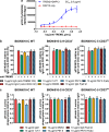
Activation of endogenous CD33 in iPSdMiG by CD33 agonistic antibodies. (a) pSYK analysis in TREM2 + DAP12 reporter cells. Addition of anti-TREM2 antibody AF1828 resulted in an increase in endogenous pSYK levels measured 30 min after the treatment only in TREM2 + DAP12 but not DAP12 expressing control reporter cells. Data are presented mean ± SD. (b) CD33 antibodies P67.6 and 1c7/1 were able to decrease the increased pSYK/tSYK levels triggered by TREM2 activation in WT iPSdMiG (left). In CD33−/− (middle) and CD33ΔE2 (right) iPSdMiG none of the tested antibodies was able to modulate pSYK/tSYK levels after TREM2 activation. Data are presented mean + SEM; n = 3–6; ** p ≤ 0.01 compared to IgG1 (anti-CD33 Ctrl) determined by Welch ANOVA followed by Games-Howell post hoc test. (c) CD33 antibodies P67.6 and 1c7/1 decreased the phagocytic uptake of pHrodo S. aureus BioParticles in WT iPSdMiG (left). In CD33−/− (middle) and CD33ΔE2 (right) iPSdMiG none of the tested antibodies was able to modulate pHrodo S. aureus BioParticle phagocytosis. Data are presented mean + SEM; n = 3; ** p ≤ 0.01 and * p ≤ 0.05 compared to IgG1 determined by ANOVA followed by Dunnett’s post hoc test.

References

    1. Brinkman-Van der Linden EC, Angata T, Reynolds SA, Powell LD, Hedrick SM, Varki A. CD33/Siglec-3 binding specificity, expression pattern, and consequences of gene deletion in mice. Mol. Cell Biol. 2003;23(12):4199–4206. doi: 10.1128/MCB.23.12.4199-4206.2003. - DOI - PMC - PubMed
    1. Crocker PR, Paulson JC, Varki A. Siglecs and their roles in the immune system. Nat. Rev. Immunol. 2007;7(4):255–266. doi: 10.1038/nri2056. - DOI - PubMed
    1. Griciuc A, Serrano-Pozo A, Parrado AR, Lesinski AN, Asselin CN, Mullin K, Hooli B, Choi SH, Hyman BT, Tanzi RE. Alzheimer's disease risk gene CD33 inhibits microglial uptake of amyloid beta. Neuron. 2013;78(4):631–643. doi: 10.1016/j.neuron.2013.04.014. - DOI - PMC - PubMed
    1. Malik M, Simpson JF, Parikh I, Wilfred BR, Fardo DW, Nelson PT, Estus S. CD33 Alzheimer’s risk-altering polymorphism, CD33 expression, and exon 2 splicing. J. Neurosci. 2013;33(33):13320–13325. doi: 10.1523/JNEUROSCI.1224-13.2013. - DOI - PMC - PubMed
    1. Siddiqui SS, Springer SA, Verhagen A, Sundaramurthy V, Alisson-Silva F, Jiang W, Ghosh P, Varki A. The Alzheimer’s disease-protective CD33 splice variant mediates adaptive loss of function via diversion to an intracellular pool. J. Biol. Chem. 2017;292(37):15312–15320. doi: 10.1074/jbc.M117.799346. - DOI - PMC - PubMed

Publication types

MeSH terms