. 2021 Jun 29;11(1):13516.
doi: 10.1038/s41598-021-92878-6.

Development of a bioavailable Hg(II) sensing system based on MerR-regulated visual pigment biosynthesis

Affiliations

Development of a bioavailable Hg(II) sensing system based on MerR-regulated visual pigment biosynthesis

Yan Guo et al. Sci Rep. .

Abstract

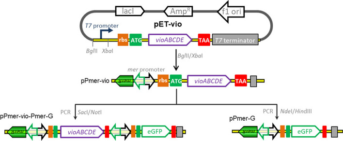
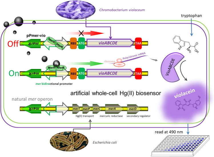
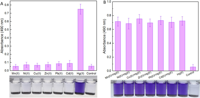
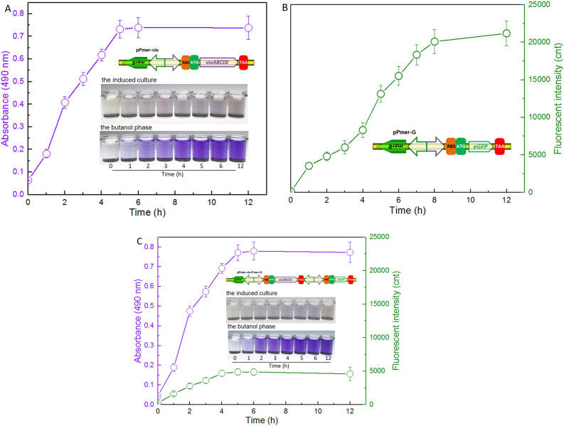
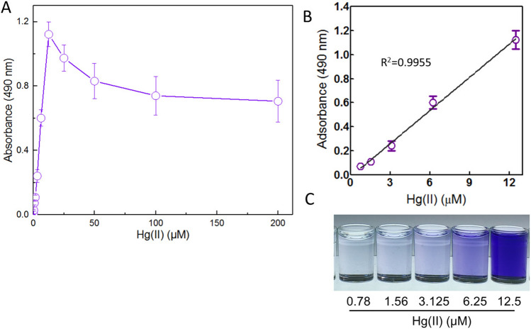
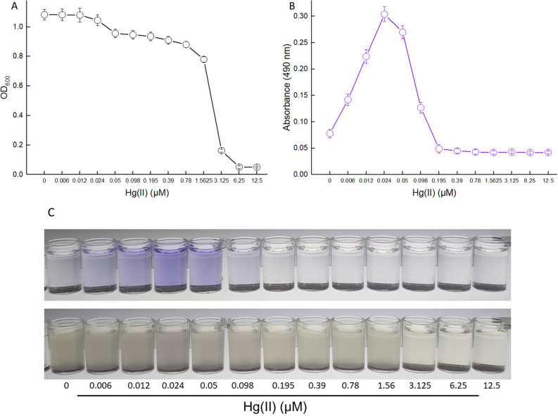
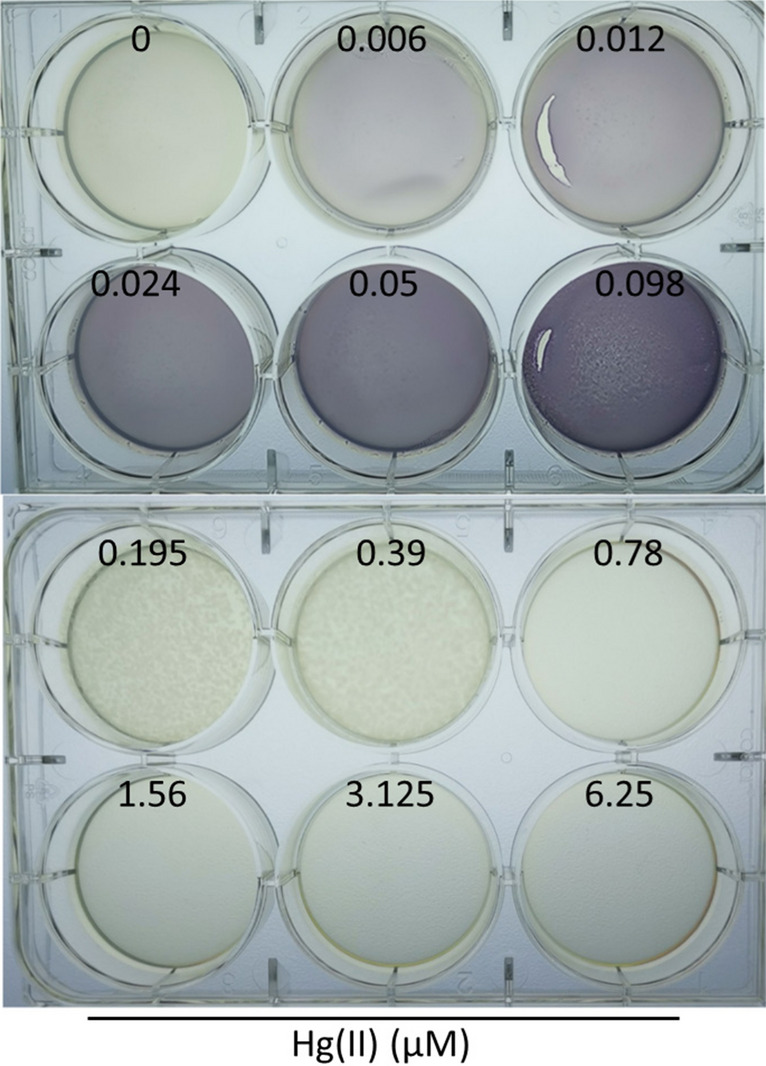
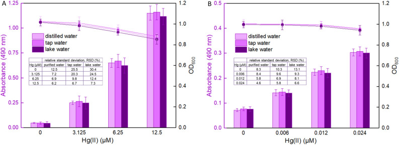
Engineered microorganisms have proven to be a highly effective and robust tool to specifically detect heavy metals in the environment. In this study, a highly specific pigment-based whole-cell biosensor has been investigated for the detection of bioavailable Hg(II) based on an artificial heavy metal resistance operon. The basic working principle of biosensors is based on the violacein biosynthesis under the control of mercury resistance (mer) promoter and mercury resistance regulator (MerR). Engineered biosensor cells have been demonstrated to selectively respond to Hg(II), and the specific response was not influenced by interfering metal ions. The response of violacein could be recognized by the naked eye, and the time required for the maximum response of violacein (5 h) was less than that of enhanced green fluorescence protein (eGFP) (8 h) in the single-signal output constructs. The response of violacein was almost unaffected by the eGFP in a double-promoter controlled dual-signals output construct. However, the response strength of eGFP was significantly decreased in this genetic construct. Exponentially growing violacein-based biosensor detected concentrations as low as 0.39 μM Hg(II) in a colorimetric method, and the linear relationship was observed in the concentration range of 0.78-12.5 μM. Non-growing biosensor cells responded to concentrations as low as 0.006 μM Hg(II) in a colorimetric method and in a Hg(II) containing plate sensitive assay, and the linear relationship was demonstrated in a very narrow concentration range. The developed biosensor was finally validated for the detection of spiked bioavailable Hg(II) in environmental water samples.


Conflict of interest statement

The authors declare no competing interests.

Figures

Figure 1
Figure 1
Assembly of artificial mer operons for sensing of bioavailable Hg(II). The violacein biosynthesis module and fluorescent reporter module were placed under the control of the mer promoter separately or in combination using genetic methods.
Figure 2
Figure 2
Engineered Hg(II) whole-cell biosensor based on MerR-regulated visual violacein production. The archetype of natural mer operon originated from E. coli is shown at the bottom of the figure. To assemble Hg(II)-responsive visual biosensor, the mercury detoxification gene cluster merTPCAD was substituted with the violacein biosynthetic gene cluster vioABCDE. The violacein biosynthetic module, originated from Chromobacterium violaceum, is transcriptionally regulated by the Hg(II) sensing element to enable a whole-cell violacein-based Hg(II) biosensor. The violacein, produced with bioavailable Hg(II) induction, can be extracted with organic solvents such as butanol, and quantified by a microplate reader at 490 nm.
Figure 3
Figure 3
The biodetection selectivity of engineered whole-cell Hg(II) biosensor. Biosensor cells in the exponential growth phase were exposed to 8 μM of various metal ions alone (A) or in combination (B) at 37 °C for 12 h. The control groups were not supplemented with metal ions. The butanol phases containing violacein were prepared and read at 490 nm in a microplate reader. The photo shown at the bottom is representative of three independent experiments with similar results. The absorbance values of pigment were normalized to bacterial cell density at 600 nm. The results are shown as the mean of three independent assays ± the standard deviation.
Figure 4
Figure 4
Time courses of reporter signals generated by three whole-cell biosensors with 8 μM Hg(II) exposure. Quantitative time-course profiles of whole-cell biosensors TOP10/pPmer-vio (A), TOP10/pPmer-G (B), and TOP10/pPmer-vio-Pmer-G (C) to Hg(II). Three kinds of bacterial cells in exponential growth phase were exposed to 8 μM Hg(II) at 37 oC. The fluorescent signal and pigment-based color change were detected at regular time intervals. Both of the reporter signals were normalized to bacterial cell density at 600 nm. The results are shown as the mean of three independent assays ± the standard deviation.
Figure 5
Figure 5
The response of exponential-phase culture of TOP10/pPmer-vio induced with increased concentrations of Hg(II). Exponential-phase culture of TOP10/pPmer-vio was induced with increased concentration of Hg(II) at 37 °C for 5 h. Whole-cell biosensor dose–response (A) and linear response (B) to Hg(II). The butanol phases containing violacein with 0.78–12.5 μM Hg(II) induction (C). Shown is one representative of three independent experiments with similar results. The background intensity (the absorbance at 490 nm with no Hg(II) exposure) was subtracted from each reading. The results are shown as the mean of three independent assays ± the standard deviation. The linear relationship was in the concentration range of 0.78–12.5 μM (R2 = 0.9955).
Figure 6
Figure 6
The response of resting TOP10/pPmer-vio induced with increased concentrations of Hg(II). E. coli TOP10 harboring the plasmid pPmer-vio was inoculated in LB broth containing increased concentrations of Hg(II) at 37 °C for 12 h. The bacterial density (OD600) was determined (A), and then the violacein was extracted and quantified at 490 nm (B). The induced culture and the butanol phase with increased concentrations of Hg(II) exposure (C). Shown is one representative of three independent experiments with similar results.
Figure 7
Figure 7
The detection sensitivity of TOP10/pPmer-vio toward Hg(II) on the LB-agar plate. Exponential-phase culture of TOP10/pPmer-vio was spread on LB-agar plates with increasing concentrations of Hg(II), and cultured at 37 °C overnight. A representative image of three independent experiments with similar results is shown.
Figure 8
Figure 8
Performance of biosensor cells against environmental water samples spiked with different concentrations of Hg(II). LB-incubated exponential-phase cultures (A) or LB-incubated lag-phase cultures (B) of whole-cell biosensor TOP10/pPmer-vio were exposed to varying concentrations of Hg(II) in the following water samples: sterile distilled water, unsterilized tap water and unsterilized lake water. After culturing at 37 °C for 12 h, the bacterial density (OD600) was first determined (point line diagram, right-Y scale), and then the butanol-extracted violacein was determined at 490 nm (bar chart, left-Y scale). Relative standard deviation (RSD) is shown in the table embodied in the figure.

References

    1. Bernhoft, R.A. Mercury toxicity and treatment: a review of the literature. J Environ Public Health2012, 460508 (2012). - PMC - PubMed
    1. Regnell O, Watras CJ. Microbial mercury methylation in aquatic environments: a critical review of published field and laboratory studies. Environ. Sci. Technol. 2019;53:4–19. doi: 10.1021/acs.est.8b02709. - DOI - PubMed
    1. Suvarapu LN, Baek SO. Recent studies on the speciation and determination of mercury in different environmental matrices using various analytical techniques. Int. J. Anal. Chem. 2017;2017:3624015. doi: 10.1155/2017/3624015. - DOI - PMC - PubMed
    1. Bereza-Malcolm LT, Mann G, Franks AE. Environmental sensing of heavy metals through whole cell microbial biosensors: a synthetic biology approach. ACS Synth. Biol. 2015;4:535–546. doi: 10.1021/sb500286r. - DOI - PubMed
    1. Jung J, Lee SJ. Biochemical and biodiversity insights into heavy metal ion-responsive transcription regulators for synthetic biological heavy metal sensors. J. Microbiol. Biotechnol. 2019;29:1522–1542. doi: 10.4014/jmb.1908.08002. - DOI - PubMed

Publication types

MeSH terms