

Case Reports
doi: 10.4103/ijd.IJD_534_19.
Dermoscopic Features of Leukemia Cutis-Case Series
Affiliations
- PMID: 34188276
- PMCID: PMC8208274
- DOI: 10.4103/ijd.IJD_534_19
Item in Clipboard
Case Reports
Dermoscopic Features of Leukemia Cutis-Case Series
Indian J Dermatol.
2021 Mar-Apr.
Abstract
Leukemia cutis (LC) is a term describing skin lesions caused by cutaneous infiltration by hematological malignancies (myeloid or lymphoid). To our knowledge, there are no published reports on dermoscopic presentation of LC. The aim of the study was to analyze dermoscopic pattern in series of 5 patients with the diagnosis of LC.
Keywords: Chloroma; dermatoscopy; dermosopy; leukemia cutis; myeloid sarcoma.
Copyright: © 2021 Indian Journal of Dermatology.
Conflict of interest statement
There are no conflicts of interest.
Figures
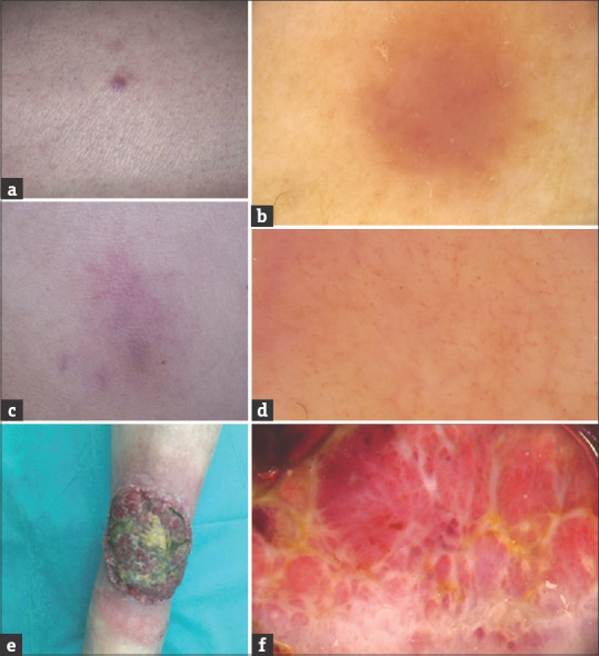
(a and b) LC in the course of acute monocytic leukemia (Patient 1). Single brownish tumor of the right lower leg (a); Dermoscopy shows diffuse pink–brownish structureless area (b). (c and d) LC in the course of chronic myelomonocytic leukemia (Patient 2). Violaceous plaque in the abdominal region (c); Dermoscopy shows polymorphic vascular pattern with the presence of dotted vessels, linear curved vessels, and linear vessels with branches as well as subtle structureless yellowish-orange areas in patchy distribution (d). (e and f) LC in the course of chronic lymphocytic leukemia (Patient 3). Giant tumor of the left lower leg (e); Dermoscopy shows vascular areas containing polymorphic vascular pattern with the presence of dotted vessels, linear curved vessels, and linear vessels with branches on the pink-red structureless background intersected with white thick lines arranged in network-like structure and yellow-red structureless areas corresponding with the presence of a crust (f)
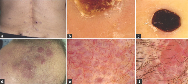
(a-c) LC in the course of chronic myelomonocytic leukemia (Patient 4). Multiple pink-brownish tumors, some with central necrotic crust, in the lumbar region (a); Dermoscopy shows the presence of polymorphic vessels on the whitish structureless background which surround central yellow-red structureless area corresponding with the presence of a crust (b). Within the lesions with central necrotic crust (black structureless area), peripheral vascular pattern was absent (c). (d-f) LC in the course of chronic lymphocytic leukemia (Patient 5). Coalescing tumors and plaques of the parietal region of the scalp (d); Dermoscopy shows the presence of polymorphic vascular pattern with the predominance of linear vessels with branches of large diameter on the background consisting of pink, round structureless areas intersected with white structureless areas; additionally subtle structureless yellowish-orange areas in random distribution may be observed (e and f)
References
-
- Wagner G, Fenchel K, Back W, Schulz A, Sachse MM. Leukemia cutis - epidemiology, clinical presentation, and differential diagnoses. J Dtsch Dermatol Ges. 2012;10:27–36. - PubMed
-
- Cho-Vega JH, Medeiros LJ, Prieto VG, Vega F. Leukemia cutis. Am J Clin Pathol. 2008;129:130–42. - PubMed
-
- Errichetti E, Zalaudek I, Kittler H, Apalla Z, Argenziano G, Bakos R, et al. Standardization of dermoscopic terminology and basic dermoscopic parameters to evaluate in general dermatology (non-neoplastic dermatoses): An expert consensus on behalf of the International dermoscopy society. Br J Dermatol. 2020;182:454–67. - PubMed
-
- Su WP. Clinical, histopathologic, and immunohistochemical correlations in leukemia cutis. Semin Dermatol. 1994;13:223–30. - PubMed
Publication types
LinkOut - more resources
Full Text Sources