Review
. 2021 May 10;52(2):e4044683.
doi: 10.25100/cm.v52i2.4683.

Damage control surgery in lung trauma

Affiliations
Review

Damage control surgery in lung trauma

Alberto García et al. Colomb Med (Cali). .

Abstract

Damage control techniques applied to the management of thoracic injuries have evolved over the last 15 years. Despite the limited number of publications, information is sufficient to scatter some fears and establish management principles. The severity of the anatomical injury justifies the procedure of damage control in only few selected cases. In most cases, the magnitude of the physiological derangement and the presence of other sources of bleeding within the thoracic cavity or in other body compartments constitutes the indication for the abbreviated procedure. The classification of lung injuries as peripheral, transfixing, and central or multiple, provides a guideline for the transient bleeding control and for the definitive management of the injury: pneumorraphy, wedge resection, tractotomy or anatomical resection, respectively. Identification of specific patterns such as the need for resuscitative thoracotomy, or aortic occlusion, the existence of massive hemothorax, a central lung injury, a tracheobronchial injury, a major vascular injury, multiple bleeding sites as well as the recognition of hypothermia, acidosis or coagulopathy, constitute the indication for a damage control thoracotomy. In these cases, the surgeon executes an abbreviated procedure with packing of the bleeding surfaces, primary management with packing of some selected peripheral or transfixing lung injuries, and the postponement of lung resection, clamping of the pulmonary hilum in the most selective way possible. The abbreviation of the thoracotomy closure is achieved by suturing the skin over the wound packed, or by installing a vacuum system. The management of the patient in the intensive care unit will allow identification of those who require urgent reintervention and the correction of the physiological derangement in the remaining patients for their scheduled reintervention and definitive management.

Las técnicas de control de daños aplicadas al manejo de lesiones torácicas han evolucionado en los últimos 15 años. A pesar de que el número de publicaciones es limitado, la información es suficiente para desvirtuar algunos temores y establecer los principios de manejo. La severidad del compromiso anatómico justifica el procedimiento de control de daños solamente en algunos casos. En la mayoría, la magnitud del deterioro fisiológico y la presencia de otras fuentes de sangrado dentro del tórax o en otros compartimientos corporales constituyen la indicación del procedimiento abreviado. La clasificación de la lesión pulmonar como periférica, transfixiante y central o múltiple, proporciona una pauta para el control transitorio del sangrado y para el manejo definitivo de la lesión: neumorrafía, resección en cuña, tractotomía o resecciones anatómicas, respectivamente. La identificación de ciertos patrones como la necesidad de toracotomía de reanimación o de oclusión aórtica, la existencia de un hemotórax masivo, de una lesión pulmonar central, una lesión traqueobronquial o una lesión vascular mayor, así como el reconocimiento de hipotermia, acidosis o coagulopatía, constituyen la indicación de una toracotomía de control de daños. En estos casos, el cirujano concluye de manera abreviada los procedimientos con empaquetamiento de las superficies sangrantes, el manejo primario con empaquetamiento de algunas lesiones pulmonares periféricas o transfixiante seleccionadas y el aplazamiento de la resección pulmonar, pinzando el hilio de la manera más selectiva posible. La abreviación del cierre de la toracotomía se logra con la sutura de la piel sobre el empaquetamiento de la herida, o mediante la instalación de un sistema de presión negativa. El manejo del paciente en cuidados intensivos permitirá identificar aquellos que requieren reintervención urgente y corregir la alteración fisiológica de los restantes para su reoperación programada y manejo definitivo.

Keywords: Damage control surgery; balloon occlusion; cardiac output; chest tubes; heart arrest; hemostasis; hemostatics; hemothorax; lung injury; pneumonectomy; sternotomy; thoracotomy.


Conflict of interest statement

Conflict of Interest: The authors declare that they have no conflict of interest.

Figures

Figure 1
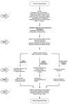
Figure 1. Algorithm for Surgical Management of Lung Trauma. Anotations: 1. Aortic occlusion can be made during the preoperative phrase by an endovascular balloon occlusion (REBOA). If the device or the protocols are not available, a left thoracotomy for aortic-cross clamping is indicated. 2. If a resuscitative thoracotomy is required, a left anterolateral incision must be performed. A sternotomy is indicated in central wounds. 3. Consider autotransfusion. 4. Packing around a peripheral injury or packing of a through- and-through wound should only be considered in patients with other more complex injuries requiring surgical team’s attention. 5. Packing lung wound should be only deemed the definitive method only in complicated cases with exhausted physiologic reserve and hemorrhage control. 6. Anatomic resection in the index operation is recommended only if hemorrhage control is immediate and physiologic is almost completely restored. 7. Temporary chest closing can be achieved by packing the wound and suturing the skin over the pads or by a vacuum system.
Figure 2
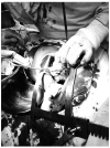
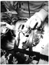
Figure 2. Transient hemorrhage control for peripheral injuries. Direct pressure with the surgeon's hand or a Duvall clamp frequently stops the hemorrhage transiently in a peripheral parenchymal wound.
Figure 3
Figure 3. Packing of the tract in a through-and- through peripheral lung wound. a) The surgeon introduces a finger gently in the tract, exploring it, and preparing to guide a hemostatic clamp. B) The hemostatic clamp has been introduced in the wound guided by the surgeon’s finger. C)The surgeon exerts traction with a laparotomy pad driving it into the wound, and gently adapting it to the wound shape. D) A Postoperative Chest X-Ray obtained immediately post procedure depicts, the intraparenchymal packing filling the lower part of the thorax, permitting the lung expansion without mediastinal deviation.
Figure 4
Figure 4. Cross clamping of the lung hilum. This patient has multiple exsanguinating lung wounds. The surgeon's left hand is introduced between the lung and the heart, making room for the clamp application. The index and middle fingers embrace the hilum to make facilitate the placement of the atraumatic clamp.
Figure 5
Figure 5. Pulmonary tractotomy. A) Two atraumatic clamps take the bridge of tissue across the tract created by the penetrating injury. The tissue is cut with a scalpel or a scissor, exposing the interior of the wound. B) The surgeon can drive the linear staple along opening of the tract, thus reducing surgical trauma, saving time, and avoiding blood loss. C) After exposing the inner aspect of the wound, the bleeding vessels and the air leaks are selectively ligated with 3/0 monofilament in figure of “8” stitches. D) If the tract was opened with clamps and scissors or scalpel, the tissue beneath the clamps must be oversewn with a monofilament 3-0 suture. Occasionally a second suture line must be performed to achieve the hemostasis, as is shown in the figure.
Figure 6
Figure 6. Clamping of the pulmonary hilum for a deferred anatomic resection. A) This patient was submitted to a resuscitative thoracotomy because of exsanguinating injuries in the lower left lobe, near the hilum, and the left hepatic lobe. He received open cardiac massage, aortic cross-clamping, and defibrillation (#3). Hemorrhage was arrested by clamping the vascular structures of the inferior lobe of the left lung and by liver packing. The chest was closed by suturing the skin over pads and the abdomen with a vacuum system. B) Postoperative chest x-ray showing the left superior lobe completely expanded. The clamp occludes the inferior left lobe's vascular structures, and there are pads packed in the lower part of the chest and upper abdomen. C) Chest X-rays after the lobectomy, which was performed two days after the index surgery.
Figure 7
Figure 7. Packing and abbreviated closure of the thoracic wall. A) This technique expedites the closure of the thoracic incision. After checking for mechanical bleeding, the surgeon places two or three laparotomy pads in the wound, between the muscular layers and the subcutaneous fat, packing the edges and by doing this, oozing around the edges of the wound is controlled. B) The skin is sutured over the pads, completing the packing effect.
Figura 1
Figura 1. Flujograma del manejo quirúrgico del trauma pulmonar. Anotaciones: 1. Durante el preoperatorio la oclusión aórtica puede hacerse mediante un catéter de oclusión intraaórtica (REBOA). Si no se dispone de este dispositivo se indica una toracotomía izquierda con este fin. 2. La esternotomía mediana está indicada en las lesiones centrales. Los pacientes que requieren toracotomía de reanimación deben ser accedidos por toracotomía anterolateral izquierda. 3. Considere autotransfusión. 4. El empaquetamiento alrededor de una herida periférica o en el tracto de una herida transfixiante solamente se debe considerar en pacientes con otras heridas más complejas qué requieren la atención del equipo quirúrgico. 5. El empaquetamiento cómo manejo definitivo de la herida pulmonar solamente se debe considerar en casos complejos en los que la reserva fisiológica se ha comprometido y se comprueba que el empaquetamiento controla la hemorragia. 6. Una resección anatómica sólo se recomienda en casos en los que se efectúa el control inmediato de la hemorragia y la estabilización es evidente con mejoría de la hipotermia y la acidosis, sin evidencia de coagulopatía.7. El cierre transitorio del tórax puede llevarse a cabo mediante empaquetamiento de la pared y sutura continua de la piel o mediante la instalación de un sistema de vacío.
Figura 2
Figura 2. Control transitorio de la hemorragia en las heridas periféricas. La compresión directa del tejido pulmonar con la mano del cirujano y con una pinza dual consiguen controlar transitoriamente el sangrado.
Figura 3
Figura 3. Empaquetamiento de una herida pulmonar transfixiante. A) El cirujano introduce el dedo índice en la herida mientras comprime el tejido pulmonar para limitar la hemorragia. Este dedo servirá de guía para el avance de una pinza. B) Una pinza hemostática ha sido insertada en la herida con la guía del dedo. C) El cirujano tracciona la compresa dentro del tracto de la herida, adaptándola a la configuración del tracto mediante movimientos suaves. D) Radiografía de tórax en el posoperatorio inmediato. Se observa que las compresas ocupan sólo una parte del espacio pleural y no desvían las estructuras mediastinales.
Figura 4
Figura 4. Pinzamiento del hilio pulmonar. Se trata de un paciente exanguinado por heridas pulmonares múltiples. El cirujano introduce su mano izquierda entre el corazón y el pulmón, creando el espacio para la introducción de la pinza. Abraza el hilio pulmonar con sus dedos índice y medio ampliando un poco más la distancia para permitir el avance de la pinza vascular con la que ocluirá las estructuras del hilio.
Figura 5
Figura 5. Tractotomía pulmonar. A) El cirujano toma el puente de tejido pulmonar con dos pinzas atraumáticas, preparando la tractotomía, que requerirá en este caso la sección del tejido con tijeras. B) El tejido pulmonar es tomado con una grapadora lineal. En este caso la sección será acompañada de hemostasia de la línea de sutura, reduciendo el trauma del pulmón. C) La apertura del tracto expone las estructuras vasculares y bronquiales lesionadas. El cirujano controla la pérdida sanguínea o aérea mediante puntos en X de monofilamento 3/0. D) Cuando el tracto ha sido abierto con tijera es necesario realizar una sutura hemostática con puntos en guardia griega por debajo de la pinza. Frecuentemente es necesario realizar un segundo plano de sutura continua para completar el efecto hemostático, tal como se muestra en esta figura.
Figura 6
Figura 6. Pinzamiento del hilio pulmonar para resección pulmonar anatómica diferida. A) Paciente con toracotomía de resucitación por lesiones exanguinantes del lóbulo pulmonar inferior izquierdo y el lóbulo hepático izquierdo. Respondió a pinzamiento aórtico, masaje cardíaco y desfibrilación (#6). Se logró control de la hemorragia con pinzamiento de los vasos del lóbulo pulmonar, empaquetamiento torácico y perihepático. B) Control radiológico posoperatorio. Se observa expansión completa del lóbulo superior, la pinza ocurriendo el hilio pulmonar y las compresas en el tórax y el abdomen. C) Radiografía de tórax después de la lobectomía, practicada semi-electivamente dos días después.
Figura 7
Figura 7. Empaquetamiento y cierre abreviado de la pared torácica. A) El empaquetamiento de la herida quirúrgica permite el cierre expedito de la cavidad toráxica en el contexto de control de daños. Después de comprobar la ausencia de sangrados mecánicos, el cirujano ubica dos o tres compresas en la herida, ajustándolas en los ángulos y las superficies sangrantes. B) Después de ubicar las compresas, el cierre se completa mediante la sutura continua de la piel, completando el efecto hemostático.

References

    1. Stone HH, Strom PR, Mullins RJ. Management of the Major Coagulopathy with Onset during Laparotomy. Ann Surg. 1983;197:532–535. doi: 10.1097/00000658-198305000-00005. - DOI - PMC - PubMed
    1. Rotondo MF, Schwab CW, McGonigal MD, Phillips GR, Fruchterman TM, Kauder DR. et al 'Damage control': An approach for improved survival in exsanguinating penetrating abdominal injury Trauma. 1993;35:375–383. doi: 10.1097/00005373-199309000-00008. - DOI - PubMed
    1. Roberts DJ, Ball CG, Feliciano D V, Moore EE, Ivatury RR, Lucas CE. History of the Innovation of Damage Control for Management of Trauma Patients 1902-2016. Ann Surg. 2017;265:1034–1044. doi: 10.1097/SLA.0000000000001803. - DOI - PubMed
    1. Zhao XG, Ma YF, Zhang M, Gan JX, Xu SW, Jiang GY. Comparison of the new injury severity score and the injury severity score in multiple trauma patients. Chinese J Traumatol. 2008;11:368–371. doi: 10.1016/S1008-1275(08)60074-7. - DOI - PubMed
    1. Valdez C, Sarani B, Young H, Amdur R, Dunne J, Chawla LS. Timing of death after traumatic injury-a contemporary assessment of the temporal distribution of death. J Surg Res. 2016;200:604–609. doi: 10.1016/j.jss.2015.08.031. - DOI - PubMed