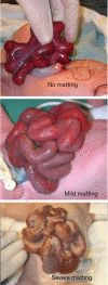
Abdominal wall defects
- PMID: 34189105
- PMCID: PMC8193006
- DOI: 10.21037/tp-20-94
Abdominal wall defects
Abstract
Abdominal wall defects are common congenital anomalies with the most frequent being gastroschisis and omphalocele. Though both are the result of errors during embryologic development of the fetal abdominal wall, gastroschisis and omphalocele represent unique disorders that have different clinical sequelae. Gastroschisis is generally a solitary anomaly with postnatal outcomes related to the underlying integrity of the prolapsed bowel. In contrast, omphalocele is frequently associated with other structural anomalies or genetic syndromes that contribute more to postnatal outcomes than the omphalocele defect itself. Despite their embryological differences, both gastroschisis and omphalocele represent anomalies of fetal development that benefit from multidisciplinary and translational approaches to care, both pre- and postnatally. While definitive management of abdominal wall defects currently remains in the postnatal realm, advancements in prenatal diagnostics and therapies may one day change that. This review focuses on recent advancements, novel techniques, and current controversies related to the prenatal diagnosis and management of gastroschisis and omphalocele.
Keywords: Abdominal wall defects; gastroschisis; omphalocele.
2021 Translational Pediatrics. All rights reserved.
Conflict of interest statement
Conflicts of Interest: Both authors have completed the ICMJE uniform disclosure form (available at http://dx.doi.org/10.21037/tp-20-94). The series “Fetal Surgery” was commissioned by the editorial office without any funding or sponsorship. The authors have no other conflicts of interest to declare.
Figures


Similar articles
-
Anomalies associated with gastroschisis and omphalocele: analysis of 2825 cases from the Texas Birth Defects Registry.J Pediatr Surg. 2014 Apr;49(4):514-9. doi: 10.1016/j.jpedsurg.2013.11.052. Epub 2013 Nov 18. J Pediatr Surg. 2014. PMID: 24726103
-
Fetal omphalocele and gastroschisis: a review of 24 cases.AJR Am J Roentgenol. 1986 Nov;147(5):1047-51. doi: 10.2214/ajr.147.5.1047. AJR Am J Roentgenol. 1986. PMID: 2945411
-
Abdominal wall defects and congenital heart disease.Ultrasound Obstet Gynecol. 2003 Apr;21(4):334-7. doi: 10.1002/uog.93. Ultrasound Obstet Gynecol. 2003. PMID: 12704739
-
Fetal abdominal wall defects.Best Pract Res Clin Obstet Gynaecol. 2014 Apr;28(3):391-402. doi: 10.1016/j.bpobgyn.2013.10.003. Epub 2013 Dec 3. Best Pract Res Clin Obstet Gynaecol. 2014. PMID: 24342556 Review.
-
Congenital abdominal wall defects and reconstruction in pediatric surgery: gastroschisis and omphalocele.Surg Clin North Am. 2012 Jun;92(3):713-27, x. doi: 10.1016/j.suc.2012.03.010. Epub 2012 Apr 17. Surg Clin North Am. 2012. PMID: 22595717 Review.
Cited by
-
Unexpected Detection of Cephalad Renal Ectopia Due to Large Omphalocele Containing the Liver on Tc-99m DMSA Scintigraphy.Mol Imaging Radionucl Ther. 2025 Feb 7;34(1):85-87. doi: 10.4274/mirt.galenos.2024.68095. Mol Imaging Radionucl Ther. 2025. PMID: 39918130 Free PMC article.
-
Challenges and lessons learnt in the management of an HIV-exposed neonate with gastroschisis in a resource-limited setting: case report.Ann Med Surg (Lond). 2024 Mar 5;86(4):2208-2213. doi: 10.1097/MS9.0000000000001924. eCollection 2024 Apr. Ann Med Surg (Lond). 2024. PMID: 38576955 Free PMC article.
-
Ultrasonographic characteristics, genetic features, and maternal and fetal outcomes in fetuses with omphalocele in China: a single tertiary center study.BMC Pregnancy Childbirth. 2023 Sep 19;23(1):679. doi: 10.1186/s12884-023-05999-3. BMC Pregnancy Childbirth. 2023. PMID: 37726736 Free PMC article.
-
The first report of gastro-thoracoschisis in a puppy born to a pomeranian bitch afflicted with dystocia.Vet Res Commun. 2025 Mar 5;49(3):128. doi: 10.1007/s11259-025-10681-4. Vet Res Commun. 2025. PMID: 40042694
-
Engineering Assembloids to Mimic Graft-Host Skeletal Muscle Interaction.Adv Healthc Mater. 2025 Jul;14(17):e2404111. doi: 10.1002/adhm.202404111. Epub 2025 May 5. Adv Healthc Mater. 2025. PMID: 40320876 Free PMC article.
References
-
- Islam S. Congenital Abdominal Wall Defects. In: Holcomb GW III, Murphy JP SPS, editor. Holcomb Ashcraft’s Pediatr. Surgery, IV. 7th ed. Philadelphia, PA: Elsevier Saunders, 2020:763-79.
Publication types
LinkOut - more resources
Full Text Sources
