. 2021 Dec 15;34(4):e0001020.
doi: 10.1128/CMR.00010-20. Epub 2021 Jun 30.

Microsporidiosis in Humans

Affiliations

Microsporidiosis in Humans

Bing Han et al. Clin Microbiol Rev. .

Abstract

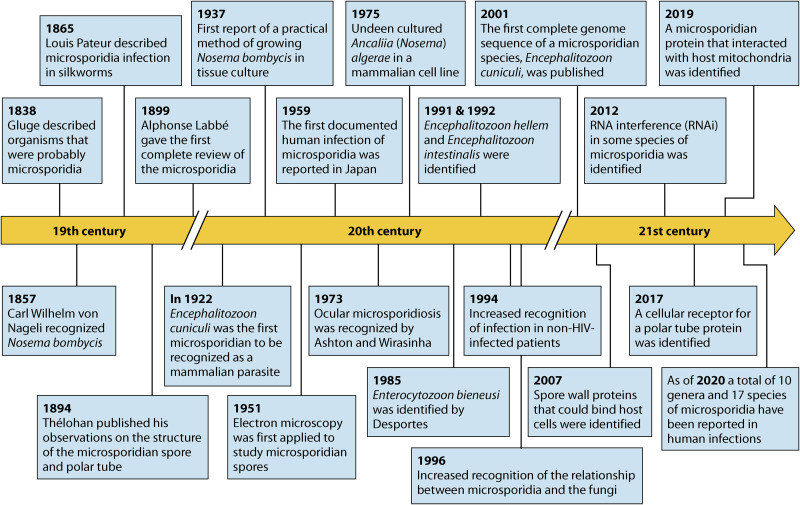
Microsporidia are obligate intracellular pathogens identified ∼150 years ago as the cause of pébrine, an economically important infection in silkworms. There are about 220 genera and 1,700 species of microsporidia, which are classified based on their ultrastructural features, developmental cycle, host-parasite relationship, and molecular analysis. Phylogenetic analysis suggests that microsporidia are related to the fungi, being grouped with the Cryptomycota as a basal branch or sister group to the fungi. Microsporidia can be transmitted by food and water and are likely zoonotic, as they parasitize a wide range of invertebrate and vertebrate hosts. Infection in humans occurs in both immunocompetent and immunodeficient hosts, e.g., in patients with organ transplantation, patients with advanced human immunodeficiency virus (HIV) infection, and patients receiving immune modulatory therapy such as anti-tumor necrosis factor alpha antibody. Clusters of infections due to latent infection in transplanted organs have also been demonstrated. Gastrointestinal infection is the most common manifestation; however, microsporidia can infect virtually any organ system, and infection has resulted in keratitis, myositis, cholecystitis, sinusitis, and encephalitis. Both albendazole and fumagillin have efficacy for the treatment of various species of microsporidia; however, albendazole has limited efficacy for the treatment of Enterocytozoon bieneusi. In addition, immune restoration can lead to resolution of infection. While the prevalence rate of microsporidiosis in patients with AIDS has fallen in the United States, due to the widespread use of combination antiretroviral therapy (cART), infection continues to occur throughout the world and is still seen in the United States in the setting of cART if a low CD4 count persists.

Keywords: AIDS; Anncaliia; Encephalitozoon; Enterocytozoon; Vittaforma; albendazole; diagnostics; fumagillin; microsporidia; prevention; therapy.


Figures

FIG 1
FIG 1
Timeline of investigations on microsporidia.
FIG 2
FIG 2
Structure of the microsporidian spore. (A) Scanning electron microscropy (SEM) image of a microsporidian spore. (B) Microsporidian spore diagram. Microsporidian spores range in size from 1 to 10 μm. The spore coat is thinner at the anterior end of the spore and consists of an electron-lucent endospore (En), an electron-dense exospore (Ex), and the plasma membrane (Pm). The sporoplasm (Sp) contains ribosomes, the posterior vacuole (PV), and a single nucleus (Nu). The anchoring disc (AD) at the anterior end of the spore is the site of attachment of the polar tube. It should be noted that the polar tube is often called the polar filament when it is within the spore prior to germination. The anterior or straight region of the polar tube that connects to the anchoring disc is called the manubroid (M), and the posterior region of the tube coils around the sporoplasm. The number of coils and their arrangement (i.e., single row or multiple rows) is used in microsporidian taxonomic classification. The lamellar polaroplast (Pl) and vesicular polaroplast (VPl) surround the manubroid region of the polar tube. The insert depicts the 5 polar tube coils in this figure in a cross section illustrating that the polar tube within the spore (i.e., polar filament) has several layers of different electron densities by electron microscopy. (Reproduced from reference with permission.)
FIG 3
FIG 3
Germinated microsporidian spores. SEM (A) and transmission electron microscopy (TEM) (B) images of a germinated spore during infection. Upon exposure to suitable environmental conditions, the polar tube (PT) rapidly discharges out of the spore (SP) and then interacts with the host cell membrane, serving as a conduit for the nucleus and sporoplasm passage into the host cell.
FIG 4
FIG 4
Developmental life cycles of the microsporidia. Spores germinate and the polar tube is extruded when they are exposed to specific environmental conditions, which vary depending on the species of microsporidia. Upon germination, the polar tube is extruded and interacts with the host cell membrane, allowing invasion (see Fig. 6 for a model of this interaction). The sporoplasm traverses the polar tube, is introduced into the host cell cytoplasm, and begins to proliferate. The morphology of the organism during the proliferative phase (which is indicated by the region above the dotted line) is used in classification systems for defining the various microsporidian genera. Relationships of the various microsporidia with their host cells during the proliferative phase are described in Table 3. On the left side of the figure, the sporoplasm is uninucleate and the microsporidian genera that are defined during the development of spores, i.e., the sporogenic phase (which is indicated by the region below the dotted line), are all uninucleate. On the right side of the figure, the sporoplasm is diplokaryotic, and all of the microsporidian genera defined during spore development have diplokaryotic developmental patterns. Regardless of whether they are uninucleate or diplokaryotic, there are three basic types of developmental forms. The first developmental pathway, as illustrated by Anncaliia spp., involves cell division by binary fission that immediately follows karyokinesis. The second type of pathway, as illustrated by some Nosema spp., involves multiple fission of elongated moniliform multinucleate cells. The third type of developmental pathway, as illustrated by Endoreticulatus spp., involves the division by plasmotomy of rounded plasmodial multinucleate cells. During the proliferative phase, cells can have one to several division cycles. Many microsporidia are either in direct contact with the host cell cytoplasm or closely abutted to the host endoplasmic reticulum; however, other microsporidia are found within a parasitophorous vacuole (e.g., Encephalitozoon spp. and probably Tetramicra spp.) or are separated from the host cell cytoplasm by a thick layer of their own secretions (e.g., Pleistophora spp.). In the sporogonic phase, this thick layer of microsporidian secretions becomes the sporophorous vesicle. During sporogony, several of the microsporidian genera, such as Enterocytozoon, Nosema, Anncaliia, Ichthyosporidium, and Tetramicra, maintain direct contact with the host cell cytoplasm. A sporophorous vesicle, illustrated by a circle around the developing sporogonial stage, is formed by the other microsporidian genera. During the developmental cycle of Thelohania and the Thelohania-like part of the Vairimorpha cycle, it should be noted that the diplokarya separate and continue their development as cells with isolated nuclei. (Adapted from reference with permission.)
FIG 5
FIG 5
Transmission electron microscopy of an Encephalitozoon hellem parasitophorous vacuole. Meronts (MR) can be seen adhering to edge of the vacuole, whereas sporonts (SP), sporoblasts (SB), and mature spores (S) detach from the vacuole membrane and move to the center of the vacuole.
FIG 6
FIG 6
Model of microsporidian (Encephalitozoon) host cell invasion. The spore wall contains spore wall proteins (SWPs) that can interact with glycosaminoglycans (GAGs) and other substances in the mucin layer (green) of the gastrointestinal track. These interactions are probably involved in germination. As the polar tube germinates, the polar tube adheres to the host surface by interactions of polar tube protein 1 (PTP1) with host cell surface mannose binding proteins (MBP). This allows the polar tube to form an invasion synapse by pushing into the host cell membrane. In the formation of the invasion synapse, interactions of PTP1 (and possibly PTP4) with the host cell membrane result in the establishment of a protected microenvironment for the extruded microsporidian sporoplasm which excludes the external environment. Within the invasion synapse, epitopes of polar tube protein 4 (PTP4) that are exposed at the tip of the polar tube interact with transferrin receptor 1 (TfR1), and possibly other host cell-interacting proteins (HCIPs), at the host cell plasma membrane, triggering signaling events. During the final steps of invasion, these various interactions lead to the formation of the invasion vacuole, which can include clathrin-mediated endocytosis as well as the involvement of host cell actin. The sporoplasm (and meront) possesses surface proteins, such as sporoplasm protein 1 (SSP1), which interact with various host cell surface proteins, tethering the sporoplasm to the plasma membrane during invasion and facilitating development of the invasion vacuole. At this early stage of infection, host mitochondria are already located around the invasion vacuole. As the vacuole completes its internalization, the sporoplasm becomes a meront and starts replicating. The meront surface interacts with the invasion vacuole membrane, forming electron-dense membrane structures that allow meront SSP1 to interact with voltage-dependent anion selective channels (VDAC) located on the outer membrane of the mitochondria. The interaction of SSP1 and VDAC appears to play a crucial role in association of host cell mitochondria with the invasion vacuole, facilitating energy acquisition from the host cell by the replicating meronts.
FIG 7
FIG 7
Light microscopy of biopsy specimens from patients with microsporidiosis. (A) Methylene blue-azure II-fuchsin stain of an intestinal biopsy specimen from a patient with Encephalitozoon intestinalis (arrow) infection. (Reproduced from reference with permission.) (B) Hematoxylin and eosin stain of muscle tissue from a patient with rheumatoid arthritis treated with antibody to TNF-α demonstrating Anncaliia algerae (arrow) myositis. (C) Tissue chromotrope stain of liver biopsy specimen from an immunodeficient mouse infected with Encephalitozoon cuniculi (arrows). (D) Steiner stain of a renal biopsy specimen demonstrating Encephalitozoon hellem (black spores) within the lumen of the renal tubule. (Reproduced from reference with permission.) (E) Hematoxylin and eosin stain of brain tissue from a rabbit with torticollis, demonstrating a microgranuloma with central necrosis due to Encephalitozoon cuniculi infection. No spores are seen in this image. (Reproduced from reference with permission.) (F) Light micrograph of intestinal villus biopsy specimen (plastic section) stained with methylene blue-azure II-fuchsin from a patient with AIDS and Enterocytozoon bieneusi infection. The arrow points to microsporidia within cells. These spores were present only on the apical epithelial surface; no spores were present on the basal surface or in the lamina propria. (Reproduced from reference with permission.)
FIG 8
FIG 8
Clinical images from patients with microsporidiosis. (A) Encephalitozoon hellem keratoconjunctivitis demonstrating punctate corneal lesions (white arrows). (B) Endoscopy of jejunal mucosa of a patient with gastrointestinal microsporidiosis due to Enterocytozoon bieneusi demonstrating fusion of the villi. (Reproduced from reference with permission.) (C) Endoscopic retrograde cholangiogram (ERCP) of a patient with HIV infection (AIDS) and sclerosing cholangitis due to Enterocytozoon bieneusi infection. The ERCP demonstrates diffuse dilations of the common bile duct with irregular walls, plus areas of narrowing and dilation (arrows) of the intrahepatic bile ducts. (Reproduced from reference with permission.)
FIG 9
FIG 9
Transmission electron micrographs of biopsy specimens from patients with microsporidiosis. (A) Duodenal epithelium from a patient with AIDS and Enterocytozoon bieneusi infection demonstrating proliferating stages (P) and late sporogonial plasmodia (Sp). The arrow points to a sloughing enterocyte containing mature spores. (Reproduced from reference with permission.) (B) Intestinal biopsy specimen from a patient with AIDS and Encephalitozoon intestinalis infection demonstrating spores within vacuoles containing a fibrillar matrix. (Reproduced from reference with permission.) (C) Enterocytozoon bieneusi spore demonstrating the characteristic finding of two rows of three cross sections of the polar tube (indicated by an arrow). (Reproduced from reference with permission.) (D) Muscle biopsy specimen from a patient with rheumatoid arthritis on an anti-TNF-α antibody who presented with myositis. Proliferative forms are seen in the biopsy specimen with a characteristic diplokaryotic nucleus (N). (E) Conjunctival biopsy specimen from a patient with keratoconjunctivitis due to Encephalitozoon hellem demonstrating characteristic spores (arrowheads) with a single row of six cross sections of the polar tube.
FIG 10
FIG 10
Demonstration of microsporidia in stool, urine, and other clinical specimens. (A and B) Chromotrope stains of stool specimens from patients with AIDS and diarrhea demonstrating spores of Enterocytozoon bieneusi measuring 0.7 to 1.0 by 1.1 to 1.6 μm. Spores have a pink to reddish hue with this stain. Stained spores can have a safety pin appearance as well. (Reproduced from reference with permission.) (C) Conjunctival scraping from a patient with Encephalitozoon hellem keratoconjunctivitis stained with a chemifluorescent brightening agent (that stains chitin) demonstrating microsporidian spores. (D) Stool specimen from a patient with AIDS and Encephalitozoon intestinalis infection stained with a quick hot Gram-chromotrope stain demonstrating spores of 1.0 to 1.2 by 2.0 to 2.5 μm. The spores have a violet hue with this stain. (Reproduced from reference with permission.) (E) Chromotrope stain of urine sediment from a patient with HIV infection who had a disseminated Encephalitozoon cuniculi infection demonstrating pink- to red-colored spores that are 1.0 to 1.5 by 2.0 to 3.0 μm. (Courtesy of Elizabeth Didier; reproduced with permission.) (F) Methylene blue-azure II-fuchsin stain of a touch preparation of an intestinal biopsy specimen (obtained by endoscopy) from a patient with intestinal Enterocytozoon bieneusi infection demonstrating intracellular spores.

References

    1. Weiss LM. 2020. Microsporidiosis, p 825–831. In Ryan ET, Hill DR, Solomon T, Aronson N, Endy TP (ed), Hunter's tropical medicine and emerging infectious diseases. Elsevier, Amsterdam, Netherlands.
    1. James TY, Pelin A, Bonen L, Ahrendt S, Sain D, Corradi N, Stajich JE. 2013. Shared signatures of parasitism and phylogenomics unite Cryptomycota and microsporidia. Curr Biol 23:1548–1553. 10.1016/j.cub.2013.06.057. - DOI - PubMed
    1. Jones MD, Forn I, Gadelha C, Egan MJ, Bass D, Massana R, Richards TA. 2011. Discovery of novel intermediate forms redefines the fungal tree of life. Nature 474:200–203. 10.1038/nature09984. - DOI - PubMed
    1. Corsaro D, Michel R, Walochnik J, Venditti D, Muller KD, Hauroder B, Wylezich C. 2016. Molecular identification of Nucleophaga terricolae sp. nov. (Rozellomycota), and new insights on the origin of the Microsporidia. Parasitol Res 115:3003–3011. 10.1007/s00436-016-5055-9. - DOI - PubMed
    1. Sprague V. 1977. Systematics of the microsporidia, p 1–510. In Bulla LA, Cheng TC (ed), Comparative pathobiology, vol 2. Plenum Press, New York, NY.

Publication types