. 2021 Dec 15;34(4):e0005019.
doi: 10.1128/CMR.00050-19. Epub 2021 Jun 30.

Evolutionary Pathways and Trajectories in Antibiotic Resistance

Affiliations

Evolutionary Pathways and Trajectories in Antibiotic Resistance

F Baquero et al. Clin Microbiol Rev. .

Abstract

Evolution is the hallmark of life. Descriptions of the evolution of microorganisms have provided a wealth of information, but knowledge regarding "what happened" has precluded a deeper understanding of "how" evolution has proceeded, as in the case of antimicrobial resistance. The difficulty in answering the "how" question lies in the multihierarchical dimensions of evolutionary processes, nested in complex networks, encompassing all units of selection, from genes to communities and ecosystems. At the simplest ontological level (as resistance genes), evolution proceeds by random (mutation and drift) and directional (natural selection) processes; however, sequential pathways of adaptive variation can occasionally be observed, and under fixed circumstances (particular fitness landscapes), evolution is predictable. At the highest level (such as that of plasmids, clones, species, microbiotas), the systems' degrees of freedom increase dramatically, related to the variable dispersal, fragmentation, relatedness, or coalescence of bacterial populations, depending on heterogeneous and changing niches and selective gradients in complex environments. Evolutionary trajectories of antibiotic resistance find their way in these changing landscapes subjected to random variations, becoming highly entropic and therefore unpredictable. However, experimental, phylogenetic, and ecogenetic analyses reveal preferential frequented paths (highways) where antibiotic resistance flows and propagates, allowing some understanding of evolutionary dynamics, modeling and designing interventions. Studies on antibiotic resistance have an applied aspect in improving individual health, One Health, and Global Health, as well as an academic value for understanding evolution. Most importantly, they have a heuristic significance as a model to reduce the negative influence of anthropogenic effects on the environment.

Keywords: antibiotic resistance; evolutionary biology; evolutionary pathways; evolutionary trajectories; pathways; trajectories.


Figures

FIG 1
FIG 1
The basic nested structure of the evolutionary units involved in antibiotic resistance. From left to right, a resistance gene is caught by a gene capture platform (as an integron), which might, in turn, be inserted into a conjugative mobile genetic element (as a plasmid), which is acquired by a particular bacterial clone. This clone is inserted in the host microbiome; the host is part of an environment where the resistance gene contributes to the environmental resistome. Evolutionary units are units of selection, i.e., they can be independently selected. The small diagram at the bottom left shows that all of these successive steps are due to internal (cellular) cis-acting transmission events (resulting in concentric rings), followed by unenclosed trans-acting transmission events (clone with resistance plasmid, host microbiota, or the environment), for example, when a bacterial cell containing a plasmid and a gene (concentric rings) is transmitted from a human host to another host and then to the environment (black line).
FIG 2
FIG 2
Units of selection as evolutionary units. A bacterial cell and a conjugative plasmid carrying antibiotic resistance genes constitute different evolutionary units, given that they are independent beneficiaries. At the top, a resistance gene that is externally acquired (small red rectangle) by the cell can be integrated either in the chromosome (black string ball) or in a conjugative plasmid (blue ring). In a selective event, the cell with the red gene in the chromosome reaches 4 copies, but the plasmid is independently transferred to a different bacterial cell (green), which is also selected and reaches 4 copies. At the end, the balance for each type of cell is 4 copies, with 8 copies for the plasmid, indicating that under this single selective antibiotic event, the plasmid is a better beneficiary than any of the bacterial cells hosting it; in other words, the plasmid is an independent unit of selection, a different evolutionary unit.
FIG 3
FIG 3
The topological interactions of bacterial populations in space and time: from clones to spinning evolutionary trajectories. Bacterial species have a complex population structure consisting of clonal ensembles linked by phylogenetic relations, which can be represented as a network in a plane (left). These clonal ensembles are sequentially maintained (left to right), but there is the possibility of clonal variation or recombination over time (red arrows). The structure of each bacterial species is frequently in the neighborhood of other species with their own structure. This vicinity is represented by a larger cylinder consisting of both of the species (middle) and enables horizontal genetic interactions (red arrows). In complex ecosystems (such as microbiotas), several cylinders are ecologically and functionally integrated, facilitating genetic exchange among apparently distant lineages (lower section). The interactive spinning of different evolutionary strands results in a single evolutionary material, which can be represented as a rope, based on vertical and horizontal interactions (red lines), giving rise to twisted common trajectories; however, the components can eventually be untwisted in changing environments (blue bidirectional arrow). The concept depicted here is that the events resulting in antibiotic resistance influence not only the trajectory of a particular clone or species in which they emerge but also the trajectories of complex bacterial ensembles.
FIG 4
FIG 4
Fitness landscapes in antibiotic resistance. At the top is an image of the classic fitness landscape metaphor developed in 1932 by Sewall Wright, where in a bidimensional plane (black) different genotypes are represented, their corresponding height in the vertical axis showing the fitness of each genotype (reproductive success) under the conditions of the landscape. Red ovals correspond to the variation (for instance, mutation) from one genotype to another one (yellow lines). Note that series of mutations (pathways) might reach low (C), medium (D), or high (A and B) fitness peaks (for instance, reaching very high MICs), but some of these pathways might have originated just by random drift (without natural selection) in the flat area of the landscape. If this landscape is crumpled as a paper ball (bottom left), peaks can go into proximity, and the genotype selected into a peak can have access to other fitness peaks (eventually resulting in genetic recombination or exchange). At the bottom right, the paper ball is deployed to illustrate the fitness landscape.
FIG 5
FIG 5
Phylogenetic networks and antibiotic resistance: trees within trees. Two separated phylogenetic networks (gray and red, either from clones, species, families) are superimposed. Inside the branches, the mobile genetic elements (MGEs) carrying antibiotic resistance genes coevolve with their hosts. Arrows represent the various events that modify the evolution of MGEs. (A) The light blue MGE introgresses (i.e., conjugates) from the gray to the red tree. (B) The recombination with the indigenous MGE (yellow) creates a new MGE variant (green), which eventually evolves within a separate branch of the red tree. (C) A variant (dark blue) of the indigenous (light blue) MGE of the gray tree emerges (mutation, internal recombination). This variant can segregate into a new branch of the gray tree (D). The purpose is to show the mixture of MGE-bacterial associations and the eventual modifications of their coevolution, giving rise to novel MGEs able to colonize other bacterial branches.
FIG 6
FIG 6
Interactions between evolutionary networks. The top and bottom horizontal fields depict networks where the respective bacterial clones and mobile genetic elements (MGEs) harboring resistance genes evolve independently. In each of the network planes, there are selection events, amplifying the clones or MGEs (cones). Occasionally, a successful plasmid interacts with a successful clone (two-headed arrows), eventually creating a high-risk resistant clone. (Inspired by and adapted from the classic figure by Feil and Spratt [633] with permission of Annual Reviews, Inc.)
FIG 7
FIG 7
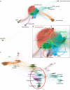
The flow of resistance genes among bacterial species should correspond to the flow of accessory genes. Shown are bipartite networks illustrating the accessory gene (protein) flow among species (genera) of the major taxa of Gammaproteobacteria (A and B) and among Firmicutes (C). Connections between two bacterial species indicate that the same accessory gene is shared, and the distance between the species (genera, in italics) is proportional to the number of connections. (B) Detail of the “core” of Enterobacteriaceae species sharing accessory genes; trumpet-like patterns on the surface of some clusters correspond to accessory genes that are unique for a particular strain (not connected with any other). The colored circles in panel B indicate the blurred borders of the species more frequently sharing accessory (and resistance) genes in Gammaproteobacteria and the “core” group exchanging accessory (and resistance) genes in Firmicutes.
FIG 8
FIG 8
Populational (clonal) fluctuations and antibiotic resistance. (A) The equilibrium between the red and blue subpopulations is locally perturbed, occasionally due to the local antibiotic selection of a recently acquired resistance trait (or a local adaptive advantage), giving rise to wave dynamics recalling a Turing instability (634). The local selection of the red population influences the blue one, which might start competing with the red, creating an expansion of instability, giving rise to new fluctuations in the equilibrium of both the red and blue populations. (B) In the upper box, three populations or clones (colored red, yellow, and gray) fluctuate in a given environment (as the microbiota). Eventually the yellow population is selected, altering the other populations. (C) The simultaneous selection of the blue and red waves results in a merging, with the emergence of a new and predominant population, a superclone (635), as might occur in environments exposed to a variety of antibiotics. At the bottom, a real fluctuation pattern of E. coli clones along years in a single hospital (unpublished data). The main concept represented here is that antibiotics contribute to the instability of the clonal structure of bacterial populations, giving rise to dominant waves that can spread across the environment.
FIG 9
FIG 9
Evolutionary pathways and trajectories. On the left, adaptive pathways and trajectories are represented as parts of a multibody pendulum, with mobile rigid members hinging on each other. The rigid parts represent the pathways, formed by broken yellow elements, corresponding to series of successive events (as mutations) leading to an efficient resistance phenotype, which are predictable and reproducible to a certain extent in the laboratory. However, these rigid parts located in bacteria oscillate by moving in different environments, where they can approach and be linked by swivel joints (white circles, representing mobile genetic linkages) to other organisms. An immense number of possible trajectories are thereby created, each offering new possibilities for interaction and linkage with other rigid parts, again eventually mediated by mobile genetic elements (the ball joints). The resulting multiple pendula greatly increases the indetermination of trajectories, approximating a chaotic behavior, with diversifying kinetics (black arrows). On the right, the possibility of loop formation among the trajectories is presented, providing a certain rigidity (and thus potential predictability) to the system. Note that the rigid parts might correspond to various units of selection, organisms, supraorganisms (such as species), and suborganisms (such as plasmids), which create a highly complex evolutionary frame.

References

    1. Davies J, Davies D. 2010. Origins and evolution of antibiotic resistance. Microbiol Mol Biol Rev 74:417–433. doi: 10.1128/MMBR.00016-10. - DOI - PMC - PubMed
    1. MacLean RC, Millan AS. 2019. The evolution of antibiotic resistance. Science 365:1082–1083. doi: 10.1126/science.aax3879. - DOI - PubMed
    1. Baquero F, Blázquez J. 1997. Evolution of antibiotic resistance. Trends Ecol Evol 12:482–487. doi: 10.1016/s0169-5347(97)01223-8. - DOI - PubMed
    1. Baquero F, Canton R. 2017. Evolutionary biology of drug resistance, p 9–32. In Mayers DL, Sobel JD, Ouellette M, Kaye KS, Marchaim D (ed), Antimicrobial drug resistance, vol 1. Mechanisms of drug resistance. Springer International Publishing, New York, NY.
    1. Ariew A. 2003. Ernst Mayr’s “ultimate/proximate” distinction reconsidered and reconstructed. Biol Philos 18:553–565. doi: 10.1023/A:1025565119032. - DOI

Publication types

Substances

LinkOut - more resources