Review
. 2021 Aug;53(4):463-487.
doi: 10.1007/s10863-021-09905-4. Epub 2021 Jun 30.

TSPO protein binding partners in bacteria, animals, and plants

Affiliations
Review

TSPO protein binding partners in bacteria, animals, and plants

Carrie Hiser et al. J Bioenerg Biomembr. 2021 Aug.

Abstract

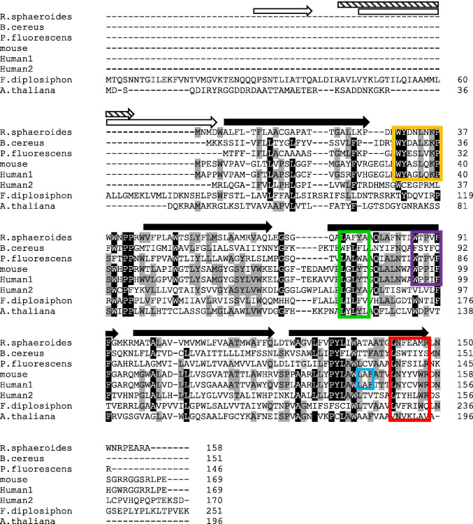
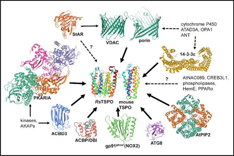
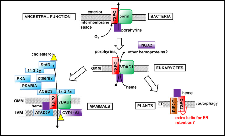
The ancient membrane protein TSPO is phylogenetically widespread from archaea and bacteria to insects, vertebrates, plants, and fungi. TSPO's primary amino acid sequence is only modestly conserved between diverse species, although its five transmembrane helical structure appears mainly conserved. Its cellular location and orientation in membranes have been reported to vary between species and tissues, with implications for potential diverse binding partners and function. Most TSPO functions relate to stress-induced changes in metabolism, but in many cases it is unclear how TSPO itself functions-whether as a receptor, a sensor, a transporter, or a translocator. Much evidence suggests that TSPO acts indirectly by association with various protein binding partners or with endogenous or exogenous ligands. In this review, we focus on proteins that have most commonly been invoked as TSPO binding partners. We suggest that TSPO was originally a bacterial receptor/stress sensor associated with porphyrin binding as its most ancestral function and that it later developed additional stress-related roles in eukaryotes as its ability to bind new partners evolved.

Keywords: 14-3-3 proteins; Autophagy; NADPH oxidase; Protein–protein interactions; TSPO; VDAC.


Conflict of interest statement

Not applicable.

Figures

Fig. 1
Fig. 1
Sequence alignment of bacterial, animal, and plant TSPO proteins. Alignment was made with Clustal Omega (Madeira et al. 2019). Black arrows indicate crystallographically-defined transmembrane helices in Rhodobacter sphaeroides TSPO (Li et al. 2016). White arrows indicate additional predicted N-terminal helices of Arabidopsis thaliana TSPO (Jurkiewicz et al. 2020). The striped arrow indicates an additional predicted N-terminal helix in Fremyella diplosiphon TSPO1 (Busch et al. 2017). The LAF and CRAC motifs are outlined in blue and red, respectively. The AIM motif (Hachez et al. 2014) is outlined in green, the 14-3-3 binding motif (Aghazadeh et al. 2012) is outlined in yellow and the WxPxF motif (Li et al. 2016) is outlined in purple. Amino acids that are 80–100% identical are shown in black with white letters; those that are 50–70% identical are shown in gray
Fig. 2
Fig. 2
TSPO protein partners. The following protein structures were obtained from the RCSB PDB and images were created using Mol* (Sehnal et al. 2018) from the PBD website: RsTSPO dimer (PDB:4UC1), mouse TSPO monomer (PDB:2NO2), human VDAC1 monomer (PDB:2JK4), Rhodobacter capsulatus porin trimer (PDB:2POR), human StAR-5 (PDB:2R55), human PKARIA (PDB:6NO7), human gp91phox (PDB:3A1F), bovine ACBP (PDB:1ACA), human ACBD3 (PDB:5LZ1), human 14-3-3ɛ (PDB:2BR9), human 14-3-3ɣ (PDB:3UZD), and yeast ATG8 (PDB:2KWC). Arabidopsis thaliana PiP2;4 (PDB:6QIM) was included as there is no available structure for AtPIP2;7
Fig. 3
Fig. 3
Evolution of TSPO functions. TSPO’s earliest function in porphyrin export from bacterial cells may have evolved into import of PPIX into and export of heme out of mitochondria and chloroplasts in eukaryotes. Functions acquired later in evolution include functioning as a SAR in plants and participating in a large protein complex that imports cholesterol into mitochondria of steroidogenic tissues in mammals. Purple squares denote porphyrins; yellow triangles denote cholesterol. Proteins involved in cholesterol transport are shaded blue; heme-containing proteins are shaded purple

References

    1. Abdrakhmanov A, Gogvadze V, Zhivotovsky B. To eat or to die: deciphering selective forms of autophagy. Trends Biochem Sci. 2020;45(4):347–364. doi: 10.1016/j.tibs.2019.11.006. - DOI - PubMed
    1. Aghazadeh Y, Papadopoulos V. The role of the 14-3-3 protein family in health, disease, and drug development. Drug Discov Today. 2016;21(2):278–287. doi: 10.1016/j.drudis.2015.09.012. - DOI - PubMed
    1. Aghazadeh Y, Rone MB, Blonder J, Ye X, Veenstra TD, Hales DB, Culty M, Papadopoulos V. Hormone-induced 14-3-3gamma adaptor protein regulates steroidogenic acute regulatory protein activity and steroid biosynthesis in MA-10 Leydig cells. J Biol Chem. 2012;287(19):15380–15394. doi: 10.1074/jbc.M112.339580. - DOI - PMC - PubMed
    1. Aghazadeh Y, Martinez-Arguelles DB, Fan J, Culty M, Papadopoulos V. Induction of androgen formation in the male by a TAT-VDAC1 fusion peptide blocking 14-3-3e protein adaptor and mitochondrial VDAC1 interactions. Mol Ther. 2014;22(10):1779–1791. doi: 10.1038/mt.2014.116. - DOI - PMC - PubMed
    1. Aghazadeh Y, Zirkin BR, Papadopoulos V. Pharmacological regulation of the cholesterol transport machinery in steroidogenic cells of the testis. Vitam Horm. 2015;98:189–227. doi: 10.1016/bs.vh.2014.12.006. - DOI - PubMed

Publication types

LinkOut - more resources