. 2021 Aug 19;32(18):1707-1723.
doi: 10.1091/mbc.E21-03-0096. Epub 2021 Jun 30.

Ventral stress fibers induce plasma membrane deformation in human fibroblasts

Affiliations

Ventral stress fibers induce plasma membrane deformation in human fibroblasts

Samuel J Ghilardi et al. Mol Biol Cell. .

Abstract

Interactions between the actin cytoskeleton and the plasma membrane are important in many eukaryotic cellular processes. During these processes, actin structures deform the cell membrane outward by applying forces parallel to the fiber's major axis (as in migration) or they deform the membrane inward by applying forces perpendicular to the fiber's major axis (as in the contractile ring during cytokinesis). Here we describe a novel actin-membrane interaction in human dermal myofibroblasts. When labeled with a cytosolic fluorophore, the myofibroblasts displayed prominent fluorescent structures on the ventral side of the cell. These structures are present in the cell membrane and colocalize with ventral actin stress fibers, suggesting that the stress fibers bend the membrane to form a "cytosolic pocket" that the fluorophores diffuse into, creating the observed structures. The existence of this pocket was confirmed by transmission electron microscopy. While dissolving the stress fibers, inhibiting fiber protein binding, or inhibiting myosin II binding of actin removed the observed pockets, modulating cellular contractility did not remove them. Taken together, our results illustrate a novel actin-membrane bending topology where the membrane is deformed outward rather than being pinched inward, resembling the topological inverse of the contractile ring found in cytokinesis.


Figures

FIGURE 1:
FIGURE 1:
Fluorescent structures are visible in HDFs loaded with a cytosolic fluorophore. After 96 h of TGF-β1 treatment, HDFs transition into myofibroblasts (Supplemental Figure S1) and separately develop fluorescent structures on the ventral side of the cell (examples marked by white arrows). These ridges can be observed in naive cells labeled with (A) cell permeable dye or cells expressing fluorescent proteins such as (B) mNeonGreen or (C) mScarlet-i. Note that, at this magnification, fluorescent puncta can be seen in cells expressing either mScarlet-i or mCherry (Figure 6) but not mNeonGreen. There is also some visible bleedthrough from the blue (Hoechst) channel into the green (Cell Explorer/mNeonGreen) channel. (Scale bar = 25 µm.) Each experiment was conducted with three technical replicates in parallel, and a representative confocal slice from one well is shown.
FIGURE 2:
FIGURE 2:
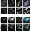
Fluorescent structures colocalize with ventral actin stress fibers. (A, B) Cells either stained with green Cell Explorer dye or constitutively expressing mNeonGreen (C, D) were fixed and stained with Hoechst (nuclei), phalloidin-California Red (actin), and an anti–phospho-paxillin primary antibody (focal adhesions) with an Alexa Fluor 647 secondary. The fluorescent structures (examples marked with white arrows) observed with either the (A) Cell Explorer dye or (C) mNeonGreen colocalize with phalloidin-stained stress fibers. The colocalized fibers have focal adhesions on both ends of the fiber (B, D), identifying them as ventral stress fibers. (Scale bar = 25 µm.) A and B and C and D are different channels for the same field of view. (Note: There is also some visible bleedthrough from the blue (Hoechst) channel into the green (Cell Explorer/mNeonGreen) channel. In addition, there is some accumulation of cytosolic fluorophore in the nucleus, as they all have a molecular weight below the 40 kDa nuclear diffusion limit [ Wei et al., 2003].) Each experiment was conducted with three technical replicates in parallel, and a representative confocal slice from one well is shown.
FIGURE 3:
FIGURE 3:
Neither invadopodial nor filopodial immunostaining colocalizes with observed fluorescent structures (A, B) HDFs stained with Hoechst (nuclei), Cell Explorer green (cytosol), Actin-Cell Mask Orange (actin), and anti-TKS5 antibody with an Alexa Fluor 647 secondary (invadopodia and podosomes). These cells have a TKS5 staining pattern similar to the negative control staining pattern provided by the manufacturer, which has no colocalization with actin stress fibers. (C, D) HDFs stained with Hoechst, Cell Explorer dye, Actin-Cell Mask Orange, and anti-Fascin antibody with an Alexa Fluor 647 secondary (filopodia) demonstrate no colocalization between the anti-Fascin antibodies and neither the actin cytoskeletion nor cytosolic pockets. (Note: There is also some visible bleedthrough from the blue (Hoechst) channel into the green (Cell Explorer) channel. In addition, there is some accumulation of cytosolic fluorophore in the nucleus, as they both have a molecular weight below the 40 kDa nuclear diffusion limit [ Wei et al., 2003].) Each experiment was conducted with three technical replicates in parallel, and a representative confocal slice from one well is shown. (Scale bar = 25 µm.)
FIGURE 4:
FIGURE 4:
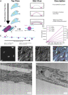
Actin stress fibers induce membrane deformation and cytosolic pocket formation, which can be visualized with cytosolic fluorophores. (A) Schematic of a proposed mechanism for the development of the fluorescent structures observed in Figure 1. As fibroblasts transition into myofibrobolasts, ventral actin stress fibers (magenta rods) originating from focal adhesions (magenta circles) deform the plasma membrane, creating cytosolic pockets (gray) for the fluorescent dye or proteins to diffuse into, leading to the observed fluorescent structures. (B) Model conceptualization of stress fiber–induced membrane deformation. The ventral stress fiber is modeled as a cylinder deforming a planar membrane. As the y dimension is uniform, the model is collapsed to one dimension. (C) Membrane deformation model used for membrane energy calculation. A stress fiber was modeled as lowering into a membrane, causing the membrane to curve. This fell into two regimes: one where the membrane is only deforming around the fiber and one where parts of the membrane beyond the fiber are deforming. The energy requirement for bending the membrane was calculated across both regimes. (D) Results from stress fiber contraction calculation. In our proposed model, the contraction of the ventral stress fibers by myosin II motors drives the formation of the cytosolic pockets. Here, we consider the energy required for that contraction over a range of observed fiber radii (50–250 nm) and contraction distances (0–15,000 nm). The calculated ATP (left y-axis) and kBT equivalents (right y-axis) indicate that the proposed model is reasonable given the timeframe of cytosolic pocket formation. (E) Fluorescent structures can be observed in the plasma membrane after staining with Green-CellBrite fixable membrane dye (examples marked with white arrows). Like the cytosolic dyes in Figure 2, these fluorescent membrane structures colocalize with ventral actin stress fibers, supporting the hypothesis that ventral actin stress fibers play a role in the formation of the observed fluorescent structures. (Scale bar = 25 µm.) (F, G) TEM images of ventral actin stress fibers where (F) membrane deformation and (G) the thick ventral stress fibers can be directly observed (white arrows). The cytosolic pockets are distinct from focal adhesions that appear as a dark plaque near the membrane and exhibit sharp curvature on the edges (Supplemental Figures S3 and S4 and [ Abercrombie et al., 1970, 1971; Medalia and Geiger, 2010]). (Scale bar = 500 nm.) Note: The CellBrite dye also brightly stains the nuclear membrane, causing some nuclear bleedthrough from the green (CellBrite) channel into the red (phalloidin) channel. Figure 5B replicates this phalloidin/membrane staining with alternate dyes and minimal bleedthrough. Each experiment was conducted with three technical replicates in parallel, and a representative confocal slice from one well is shown.
FIGURE 5:
FIGURE 5:
Direct coupling of stress fibers to the plasma membrane is necessary for membrane deformation. Immunofluorescence staining for ERM proteins reveals that ERM proteins colocalize with (A) stress fibers as well as (B) cytosolic and cell membrane pockets. (C) Before and (D) after images of cells treated with the ezrin inhibitor NSC66839. Treatment removes cytosolic pockets, but does not dissolve actin stress fibers, suggesting that physical coupling of the stress fiber to the membrane is necessary for membrane contouring. (Scale bar = 25 µm.) Each experiment was conducted with three technical replicates in parallel, and a representative confocal slice from one well is shown.
FIGURE 6:
FIGURE 6:
The structural integrity of actin stress fibers is required for membrane deformation. (A) Before and (B) after images of cells treated with cytochalasin-D. Cytochalasin-D treatment dissolved cytosolic pockets associated with stress fibers but not with focal adhesions, indicating that the physical structure of stress fibers is necessary for membrane deformation. (C) Before and (D) after images of cells treated with Blebbistatin. Blebbistatin treatment removed the observed cytosolic pockets but not the corresponding stress fibers, suggesting that once the membrane has been deformed, myosin–actin binding is necessary to maintain the cytosolic pocket. (Scale bar = 25 µm.) Each experiment was conducted with three technical replicates in parallel, and a representative confocal slice from one well is shown.
FIGURE 7:
FIGURE 7:
Active stress fiber contractility is not required to maintain membrane deformation. (A) Before and (B) after images of cells treated with calyculin A to increase cellular contractility. Calyculin A treatment did not alter the colocalization between cytosolic pockets and actin stress fibers. (C) Before and (D) after images of cells treated with Y-27632. Y-27632 treatment had no effect on the colocalization between the observed cytosolic pockets and actin stress fibers. Taken together, these two experiments suggest that once the membrane has been deformed, stress fiber contraction is not necessary to maintain membrane deformation. (Scale bar = 25 µm.) Each experiment was conducted with three technical replicates in parallel, and a representative confocal slice from one well is shown.
FIGURE 8:
FIGURE 8:
Contextualizing actin stress fiber–plasma membrane interactions. Schematics of the contractile ring in cytokinesis (left) and lateral membrane bending (right) demonstrating inverse topologies. In cytokinesis, an actin filament ring (magenta) contracts (black arrows) and applies a centripetal force (gray arrows) to the membrane (cyan), which is on the outside of the fiber, resulting in inward contraction of the membrane and ultimately membrane cleavage. In the novel ventral stress fiber–induced membrane bending, the ventral stress fibers can be viewed as an arc on a circle. In this case the membrane is on the inside of the fiber, and when these fibers contract (black arrows) and apply the centripetal force to the membrane (gray arrow), they deform the membrane and create cytosolic pockets.

Similar articles

Cited by

References

    1. Abercrombie M, Heaysman JEM, Pegrum SM (1970). The locomotion of fibroblasts in culture I. Movements of the leading edge. Exp Cell Res 59, 393–398. - PubMed
    1. Abercrombie M, Heaysman JEM, Pegrum SM (1971). The locomotion of fibroblasts in culture: IV. Electron microscopy of the leading lamella. Exp Cell Res 67, 359–367. - PubMed
    1. Amann KJ, Pollard TD (2001). Direct real-time observation of actin filament branching mediated by Arp2/3 complex using total internal reflection fluorescence microscopy. Proc Natl Acad Sci USA 98, 15009–15013. - PMC - PubMed
    1. Arpin M, Algrain M, Louvard D (1994). Membrane-actin microfilament connections: an increasing diversity of players related to band 4.1. Curr Opin Cell Biol 6, 136–141. - PubMed
    1. Baik M, French B, Chen YC, Byers JT, Chen KT, French SW, Díaz B (2019). Identification of invadopodia by TKS5 staining in human cancer lines and patient tumor samples. MethodsX 6, 718–726. - PMC - PubMed

Publication types

LinkOut - more resources