

Tempo-spatial integration of nociceptive stimuli assessed via the nociceptive withdrawal reflex in healthy humans
- PMID: 34191609
- PMCID: PMC8409952
- DOI: 10.1152/jn.00155.2021
Tempo-spatial integration of nociceptive stimuli assessed via the nociceptive withdrawal reflex in healthy humans
Abstract
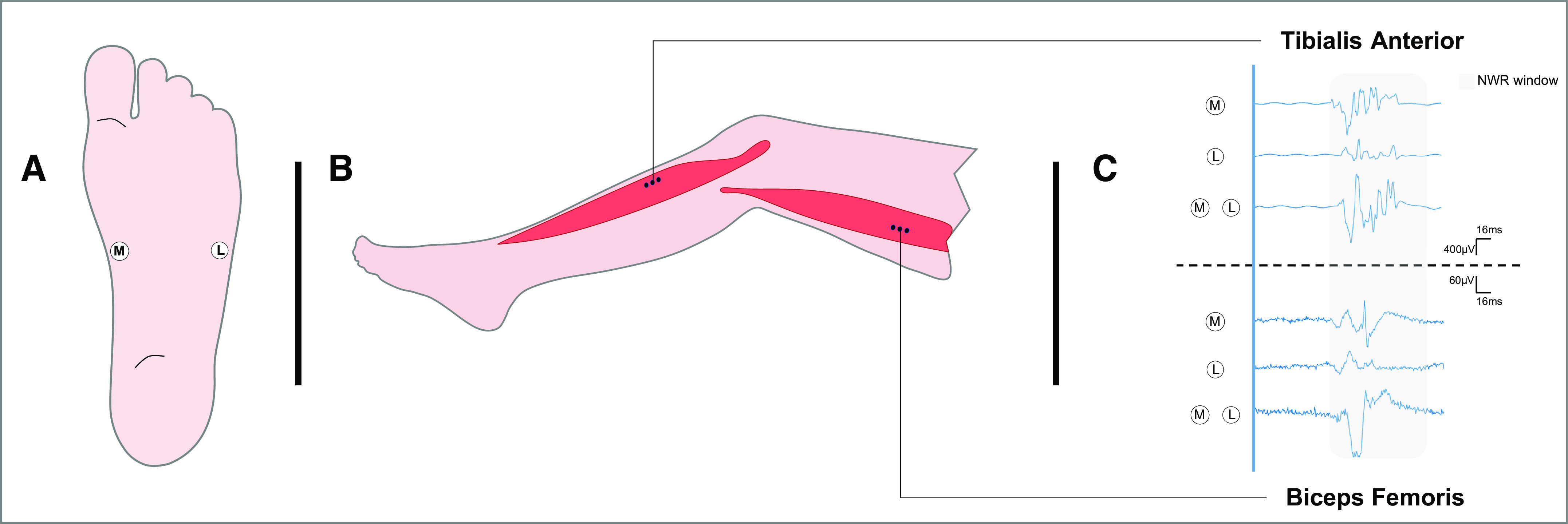
Spatial information of nociceptive stimuli applied in the skin of healthy humans is integrated in the spinal cord to determine the appropriate withdrawal reflex response. Double-simultaneous stimulus applied in different skin sites are integrated, eliciting a larger reflex response. The temporal characteristics of the stimuli also modulate the reflex, e.g., by temporal summation. The primary aim of this study was to investigate how the combined tempo-spatial aspects of two stimuli are integrated in the nociceptive system. This was investigated by delivering single- and double-simultaneous stimulation and sequential stimulation with different interstimulus intervals (ISIs ranging 30-500 ms) to the sole of the foot of 15 healthy subjects. The primary outcome measure was the size of the nociceptive withdrawal reflex (NWR) recorded from the tibialis anterior (TA) and biceps femoris (BF) muscles. Pain intensity was measured using a numerical rating scale (NRS) scale. Results showed spatial summation in both TA and BF when delivering simultaneous stimulation. Simultaneous stimulation provoked larger reflexes than sequential stimulation in TA, but not in BF. Larger ISIs elicited significantly larger reflexes in TA, whereas the opposite pattern occurred in BF. This differential modulation between proximal and distal muscles suggests the presence of spinal circuits eliciting a functional reflex response based on the specific tempo-spatial characteristics of a noxious stimulus. No modulation was observed in pain intensity ratings across ISIs. Absence of modulation in the pain intensity ratings argues for an integrative mechanism located within the spinal cord governed by a need for efficient withdrawal from a potentially harmful stimulus.NEW & NOTEWORTHY Tempo-spatial integration of electrical noxious stimuli was studied using the nociceptive withdrawal reflex and a perceived intensity. Tibialis anterior and biceps femoris muscles were differentially modulated by the temporal characteristics of the stimuli and stimulated sites. These findings suggest that spinal neurons are playing an important role in the tempo-spatial integration of nociceptive information, leading to a reflex response that is distributed across multiple spinal cord segments and governed by an efficient defensive withdrawal strategy.
Keywords: nociception; nociceptive withdrawal reflex; spatial summation; temporal summation.
Conflict of interest statement
No conflicts of interest, financial or otherwise, are declared by the authors.
Figures


Similar articles
-
Stimulus predictability moderates the withdrawal strategy in response to repetitive noxious stimulation in humans.J Neurophysiol. 2020 Jun 1;123(6):2201-2208. doi: 10.1152/jn.00028.2020. Epub 2020 Apr 29. J Neurophysiol. 2020. PMID: 32347161
-
Spinal spatial integration of nociception and its functional role assessed via the nociceptive withdrawal reflex and psychophysical measures in healthy humans.Physiol Rep. 2020 Nov;8(22):e14648. doi: 10.14814/phy2.14648. Physiol Rep. 2020. PMID: 33217191 Free PMC article.
-
Spinal Nociception is Facilitated during Cognitive Distraction.Neuroscience. 2022 May 21;491:134-145. doi: 10.1016/j.neuroscience.2022.03.038. Epub 2022 Apr 4. Neuroscience. 2022. PMID: 35381321
-
The organization of motor responses to noxious stimuli.Brain Res Brain Res Rev. 2004 Oct;46(2):163-72. doi: 10.1016/j.brainresrev.2004.07.005. Brain Res Brain Res Rev. 2004. PMID: 15464205 Review.
-
Spinal hyperexcitability in patients with chronic musculoskeletal pain or headache as evidenced by alterations in the nociceptive withdrawal reflex: a systematic review and meta-analysis.Pain. 2025 May 1;166(5):1002-1029. doi: 10.1097/j.pain.0000000000003436. Epub 2024 Oct 29. Pain. 2025. PMID: 39471047
Cited by
-
A hybrid sensory feedback system for thermal nociceptive warning and protection in prosthetic hand.Front Neurosci. 2024 Apr 8;18:1351348. doi: 10.3389/fnins.2024.1351348. eCollection 2024. Front Neurosci. 2024. PMID: 38650624 Free PMC article.
References
MeSH terms
LinkOut - more resources
Full Text Sources
Miscellaneous