. 2021 Aug 1;126(2):427-439.
doi: 10.1152/jn.00089.2021. Epub 2021 Jun 30.

BK channel activation by L-type Ca2+ channels CaV1.2 and CaV1.3 during the subthreshold phase of an action potential

Affiliations

BK channel activation by L-type Ca2+ channels CaV1.2 and CaV1.3 during the subthreshold phase of an action potential

Amber E Plante et al. J Neurophysiol. .

Abstract

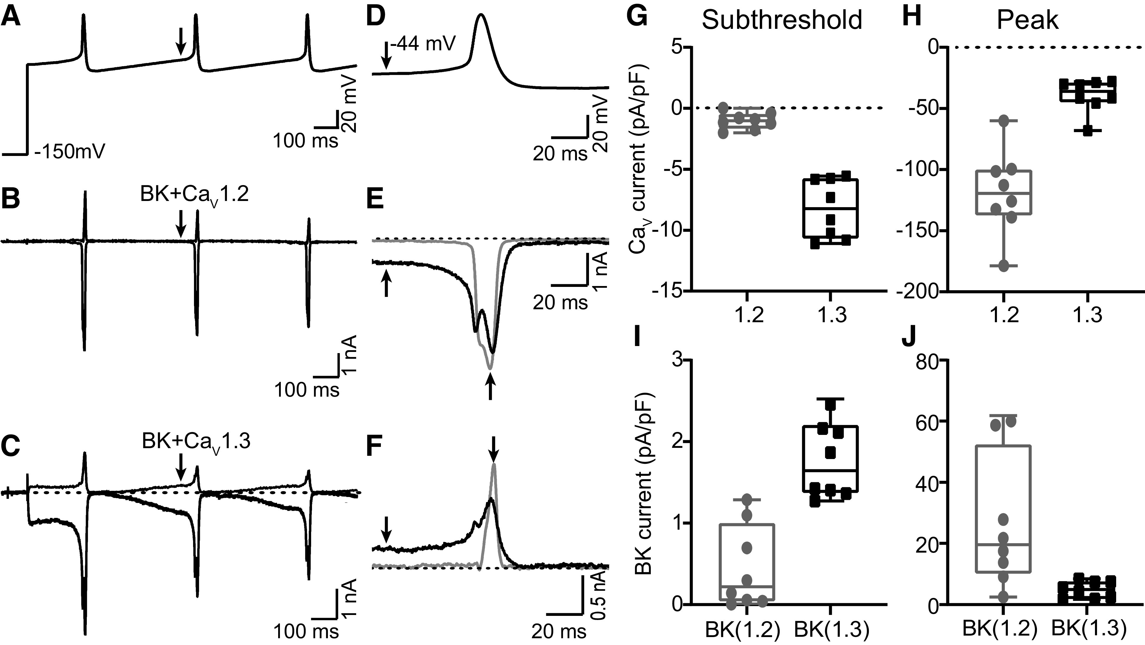
Mammalian circadian (24 h) rhythms are timed by the pattern of spontaneous action potential firing in the suprachiasmatic nucleus (SCN). This oscillation in firing is produced through circadian regulation of several membrane currents, including large-conductance Ca2+- and voltage-activated K+ (BK) and L-type Ca2+ channel (LTCC) currents. During the day steady-state BK currents depend mostly on LTCCs for activation, whereas at night they depend predominantly on ryanodine receptors (RyRs). However, the contribution of these Ca2+ channels to BK channel activation during action potential firing has not been thoroughly investigated. In this study, we used a pharmacological approach to determine that both LTCCs and RyRs contribute to the baseline membrane potential of SCN action potential waveforms, as well as action potential-evoked BK current, during the day and night, respectively. Since the baseline membrane potential is a major determinant of circadian firing rate, we focused on the LTCCs contributing to low voltage activation of BK channels during the subthreshold phase. For these experiments, two LTCC subtypes found in SCN (CaV1.2 and CaV1.3) were coexpressed with BK channels in heterologous cells, where their differential contributions could be separately measured. CaV1.3 channels produced currents that were shifted to more hyperpolarized potentials compared with CaV1.2, resulting in increased subthreshold Ca2+ and BK currents during an action potential command. These results show that although multiple Ca2+ sources in SCN can contribute to the activation of BK current during an action potential, specific BK-CaV1.3 partnerships may optimize the subthreshold BK current activation that is critical for firing rate regulation.NEW & NOTEWORTHY BK K+ channels are important regulators of firing. Although Ca2+ channels are required for their activation in excitable cells, it is not well understood how BK channels activate using these Ca2+ sources during an action potential. This study demonstrates the differences in BK current activated by CaV1.2 and CaV1.3 channels in clock neurons and heterologous cells. The results define how specific ion channel partnerships can be engaged during distinct phases of the action potential.

Keywords: BK channel; KCNMA1; L-type calcium channel; action potential; suprachiasmatic nucleus.


Conflict of interest statement

No conflicts of interest, financial or otherwise, are declared by the authors.

Figures

None
Graphical abstract
Figure 1.
Figure 1.
Voltage-gated Ca2+ channel (VGCC) inhibitor effects on daytime suprachiasmatic nucleus (SCN) action potential waveforms. A–E: paired whole cell current-clamp recordings of spontaneous action potentials obtained in wild-type (WT) SCN slices during baseline control (Ctrl) and 10 min after application of control vehicle (Veh; A) or Ca2+ channel inhibitors including 10 µM nimodipine (Nim; B), 3 µM ω-conotoxin GVIA (ConoGVIA; C), 0.3 µM ω-agatoxin IVA (AgaIVA; D), and 30 µM nickel (Ni2+; E). F and G: dantrolene (Dan) effects on daytime and nighttime action potential waveforms in WT SCN: action potentials obtained from different neurons in day (F) and night (G) under control conditions (WT) or under chronic exposure to 10 µM Dan. Dotted line indicates –50 mV.
Figure 2.
Figure 2.
Ca2+ dependence of action potential-evoked large-conductance Ca2+- and voltage-activated K+ (BK) currents in wild type (WT) and β2 knockout (β2 KO) suprachiasmatic nucleus (SCN) neurons. Prerecorded daytime SCN action potential waveforms were used as voltage commands to elicit macroscopic currents in neurons from WT and β2 KO SCN slices under control (Ctrl) conditions or chronically treated with 10 µM nimodipine (Nim) and 10 µM dantrolene (Dan) during the day and night. BK currents were isolated with 10 µM paxilline. Subthreshold (arrow) and peak action potential-evoked BK current densities were quantified from the second action potential waveform. A: daytime action potential voltage command [top; threshold, −38 mV; baseline membrane potential, −48 mV; half-width, 5.5 ms; peak, +8 mV; afterhyperpolarization (AHP), −56 mV] and BK current traces (bottom) recorded from WT SCN during the day. B and C: subthreshold (B) and peak (C) action potential-evoked BK current densities measured from WT and β2 KO SCN slices in each drug condition during the day. In WT SCN during the day, peak action potential-evoked BK current density was decreased in Nim (P = 0.01). D: nighttime action potential command (top; threshold, −43 mV; baseline membrane potential, −51 mV; half-width, 3 ms; peak, +0.9 mV; AHP, −58 mV) and BK current traces (bottom) from WT SCN during the night in Ctrl, Nim, or Dan conditions. E and F: subthreshold (E) and peak (F) action potential-evoked BK current densities measured from WT and β2 KO SCN slices in Ctrl, Nim, and Dan during the night. At night, Dan decreased subthreshold BK current in WT (P = 0.0009) and β2 KO SCN (P= 0.04). *Significance (P < 0.05) tested by 1-way ANOVA with Bonferroni’s post hoc correction comparing the drug effects for each genotype per time point. Data are means ± SE. Data points (N) are measurements from individual SCN neurons. N for WT (day, night) = Ctrl (18, 20); Nim (5, 8); Dan (7) and β2 KO (day, night) = Ctrl (7, 13), Nim (8, 12), Dan (5, 6).
Figure 3.
Figure 3.
Ca2+ and large-conductance Ca2+- and voltage-activated K+ (BK) currents from BK-CaV1.2 and BK-CaV1.3 coexpression in Chinese hamster ovary (CHO) cells. A: voltage protocol used in whole cell voltage-clamp recordings to elicit macroscopic currents from BK-CaV1.2 or BK-CaV1.3 channels. From a holding potential of −90 mV, voltages were stepped from −100 mV to +70 mV for 50 ms in 10-mV increments followed by a 100-ms step to +70 mV with 10 s between each sweep. B–D: total current (B), inward Ca2+ current (C), and outward BK K+ currents (D) from cells expressing BK and CaV1.2 channels (left) or BK and CaV1.3 channels (right). Ca2+ and BK K+ currents were isolated with 1 µM paxilline. E: current-voltage (I-V) relationships of the peak current recorded from CaV1.2, CaV1.3, BK-CaV1.2, and BK-CaV1.3 channels. Peak Ca2+ currents were measured from the 50 ms voltage steps, and peak BK currents were measured at the 100 ms +70 mV voltage step. Currents were normalized to the maximum and plotted against the 50 ms voltage step potentials. F: conductance [G, normalized to maximum (Gmax)]-voltage (V) curves for CaV1.2 and CaV1.3 currents fit to a Boltzmann function. N = 8, BK-CaV1.2; 9, BK-CaV1.3. G: I-V relationship of whole cell macroscopic Ca2+ currents from CaV1.2 and CaV1.3 channels expressed in CHO cells recorded in the presence of the BK channel α-subunit (1.2 and 1.3) or the absence of BK channel expression (1.2 no BK and 1.3 no BK). Currents were elicited with the same voltage protocol shown in A. BK channel expression had no effect on CaV1.2 or CaV1.3 currents. There were no significant differences between 1.2 and 1.2 no BK (P = 0.5) or between 1.3 and 1.3 no BK (P = 0.4) currents at any voltage step (P > 0.05, 2-way repeated-measures ANOVA. H: time constant of inactivation (tau inactivation) for CaV1.2 and CaV1.3 currents. CaV1.3 channels inactivated significantly slower than CaV1.2 between −10 and +20 mV (P = 0.02, 2-way repeated-measures ANOVA). Peak CaV1.3 currents (at −20 mV) inactivated slower than peak CaV1.2 currents (at 0 mV) (P = 0.04, unpaired Student’s t test). Data are means ± SE with N as the number of cells recorded. N = 8, 1.2; 4, 1.2 no BK; 9, 1.3; 3, 1.3 no BK.
Figure 4.
Figure 4.
Action potential-evoked large-conductance Ca2+- and voltage-activated K+ (BK) currents from BK-CaV1.2 and BK-CaV1.3 coexpression in Chinese hamster ovary (CHO) cells. A: daytime action potential voltage command (same as Fig. 2A) used to elicit macroscopic currents from BK-CaV1.2 or BK-CaV1.3 channels. From a holding potential of −90 mV, voltages were stepped to −150 mV for 100 ms, followed by a string of 3 sequential action potential waveforms. B and C: action potential-evoked currents from BK and CaV1.2 channels (B) or BK and CaV1.3 channels (C). Ca2+ and BK K+ currents were isolated with 1 µM paxilline. D: maximized view of second action potential waveform from the voltage command. E and F: representative traces of currents elicited during the second action potential waveform: overlay of Ca2+ currents from CaV1.2 and CaV1.3 channels (E) and K+ currents from BK-CaV1.2 and BK-CaV1.3 channels (F). Arrows in E and F indicate subthreshold and peak currents. G and I: box plots of median, maximum, and minimum subthreshold Ca2+ current density (G) and BK K+ current density (I). Subthreshold CaV1.3 currents were larger than CaV1.2 currents (P < 0.0001), and currents from BK-CaV1.3 channels were larger than BK-CaV1.2 currents (P = 0.0001). H and J: box plots of median, maximum, and minimum peak Ca2+ and BK K+ current densities from CaV1.2 and CaV1.3 channels (H) or BK-CaV1.2 and BK-CaV1.3 channels (J). Peak currents from CaV1.2 channels were larger than peak currents from CaV1.3 (P < 0.0001), and the peak currents from BK-CaV1.2 were larger than peak currents from BK-CaV1.3 (P = 0.01). Significance (P < 0.05) tested with unpaired Student’s t tests. N values are data points from individual cells. N = 8, 1.2; 8, 1.3; 8, BK-CaV1.2; 8, BK-CaV1.3.

Similar articles

Cited by

References

    1. Harvey JR, Plante AE, Meredith AL. Ion channels controlling circadian rhythms in suprachiasmatic nucleus excitability. Physiol Rev 100: 1415–1454, 2020. doi:10.1152/physrev.00027.2019. - DOI - PMC - PubMed
    1. Kent J, Meredith AL. BK channels regulate spontaneous action potential rhythmicity in the suprachiasmatic nucleus. PLoS One 3: e3884, 2008. doi:10.1371/journal.pone.0003884. - DOI - PMC - PubMed
    1. Meredith AL, Wiler SW, Miller BH, Takahashi JS, Fodor AA, Ruby NF, Aldrich RW. BK calcium-activated potassium channels regulate circadian behavioral rhythms and pacemaker output. Nat Neurosci 9: 1041–1049, 2006. doi:10.1038/nn1740. - DOI - PMC - PubMed
    1. Montgomery JR, Meredith AL. Genetic activation of BK currents in vivo generates bidirectional effects on neuronal excitability. Proc Natl Acad Sci USA 109: 18997–19002, 2012. doi:10.1073/pnas.1205573109. - DOI - PMC - PubMed
    1. Montgomery JR, Whitt JP, Wright BN, Lai MH, Meredith AL. Mis-expression of the BK K+ channel disrupts suprachiasmatic nucleus circuit rhythmicity and alters clock-controlled behavior. Am J Physiol Cell Physiol 304: C299–C311, 2013. doi:10.1152/ajpcell.00302.2012. - DOI - PMC - PubMed

Substances

LinkOut - more resources