

MRI characteristics of intracranial masses in the paediatric population of KwaZulu-Natal: A neuroimaging-based study
- PMID: 34192072
- PMCID: PMC8182454
- DOI: 10.4102/sajr.v25i1.2042
MRI characteristics of intracranial masses in the paediatric population of KwaZulu-Natal: A neuroimaging-based study
Abstract
Background: MRI is the imaging modality of choice for the assessment of intracranial masses in children. Imaging is vital in planning further management.
Objectives: The purpose of this study was to describe the common intracranial masses and their imaging characteristics in the paediatric population referred to Inkosi Albert Luthuli Central Hospital for MRI of the brain.
Method: We retrospectively reviewed the medical records of paediatric patients (aged from birth to 18 years) who underwent MRI investigations for intracranial masses between January 2010 and December 2016.
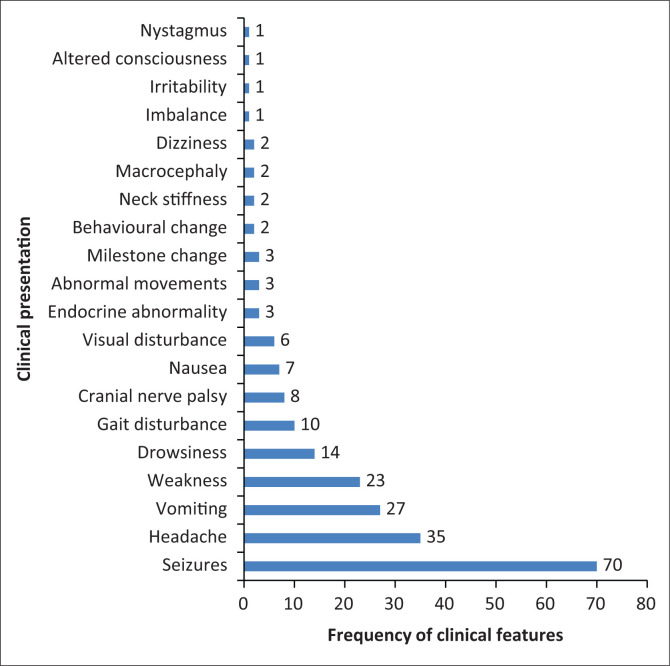
Results: A total of 931 MRI brain scans were performed. One hundred and seven scans met the inclusion criteria, of which 92 were primary brain tumours and 15 were inflammatory masses. The majority were females (56%). The mean age was 12 ± 4.52 (range of 3-18 years). The most common presenting symptom was seizures (70/107, 65.4%). We categorised the masses according to supra- and infratentorial compartments. The most common site for masses was the supratentorial compartment (n = 56, 52%). The most common masses in the supratentorial compartment were craniopharyngiomas (14/45, 31.1%), whilst in the infratentorial compartment, the most common masses were medulloblastomas (24/47, 51.1%).
Conclusion: In our series, the supratentorial compartment was the commonest site for intracranial masses. The most common tumour in the infratentorial compartment was medulloblastoma. This information is vital in formulating differential diagnoses of intracranial masses.
Keywords: brain abscess; brain tumours; intracranial masses; magnetic resonance imaging; tuberculosis.
© 2021. The Authors.
Conflict of interest statement
The authors declare that they have no financial or personal relationships that may have inappropriately influenced them in writing this article.
Figures


Similar articles
-
"A tumour registry initiative".World Neurosurg X. 2023 Jun 25;20:100227. doi: 10.1016/j.wnsx.2023.100227. eCollection 2023 Oct. World Neurosurg X. 2023. PMID: 37456693 Free PMC article.
-
Morphological pattern and frequency of intracranial tumours in children.J Coll Physicians Surg Pak. 2004 Mar;14(3):150-2. J Coll Physicians Surg Pak. 2004. PMID: 15228847
-
Neuroophthalmic Manifestations of Intracranial Tumours in Children.Case Rep Ophthalmol Med. 2021 May 15;2021:7793382. doi: 10.1155/2021/7793382. eCollection 2021. Case Rep Ophthalmol Med. 2021. PMID: 34055437 Free PMC article.
-
Paediatric brain tumours managed in Enugu, Southeast Nigeria: Review of one centre experience.Niger Postgrad Med J. 2018 Jul-Sep;25(3):186-190. doi: 10.4103/npmj.npmj_132_18. Niger Postgrad Med J. 2018. PMID: 30264771 Review.
-
Management and Outcomes of Paediatric Intracranial Suppurations in Low- and Middle-Income Countries: A Scoping Review.Front Surg. 2021 Aug 12;8:690895. doi: 10.3389/fsurg.2021.690895. eCollection 2021. Front Surg. 2021. PMID: 34466410 Free PMC article.
References
-
- Nkusi AE. Epidemiology of primary paediatric brain tumours at Johannesburg and Chris Hani Baragwanath hospitals from April 1995 to April 2005. Johannesburg: Wits Institutional Repository Environment on DSpace; 2008.
-
- Subramanian S, Ahmad T. Cancer, Childhood brain tumours. In: StatPearls. Treasure Island, FL: StatPearls Publishing; 2019. [cited 2019 Apr 11]. Available from: http://www.ncbi.nlm.nih.gov/books/NBK535415/ - PubMed
LinkOut - more resources
Full Text Sources
Miscellaneous