. 2021 Nov 1;64(11):1354-1363.
doi: 10.1097/DCR.0000000000002151.

CEACAM5, KLK6, SLC35D3, POSTN, and MUC2 mRNA Analysis Improves Detection and Allows Characterization of Tumor Cells in Lymph Nodes of Patients Who Have Colon Cancer

Affiliations

CEACAM5, KLK6, SLC35D3, POSTN, and MUC2 mRNA Analysis Improves Detection and Allows Characterization of Tumor Cells in Lymph Nodes of Patients Who Have Colon Cancer

Lina M Olsson et al. Dis Colon Rectum. .

Abstract

Background: Lymph node metastasis is the single most important prognostic risk factor for recurrence in patients with colon cancer who have undergone curative surgery. The routine method for detecting disseminated tumor cells in lymph nodes is microscopic examination of one or a few hematoxylin and eosin-stained tissue sections by a trained pathologist. This method, however, is insensitive mainly because less than 1% of the lymph node volume is examined, leading to misclassification.

Objective: This study aimed to investigate whether analysis of a selected group of biomarker mRNAs improves detection and characterization of lymph node metastases/micrometastases compared with the routine method.

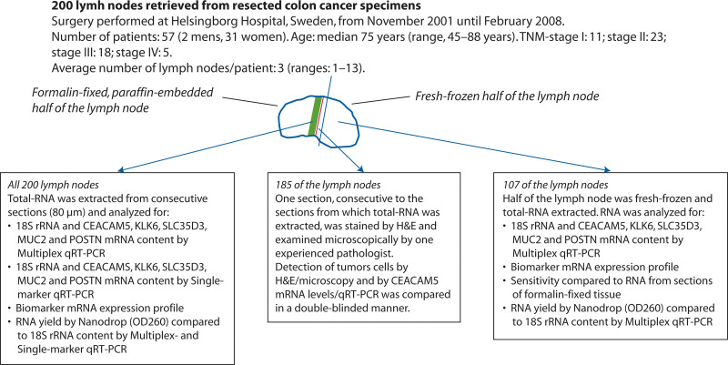
Design: This study is a side-by-side comparison of biomarker mRNA analysis and histopathology of 185 lymph nodes from patients with colon cancer representing stages I to IV, and an investigation of the importance of lymph node tissue volume for tumor cell detection.

Settings: This is a collaborative study between a high-volume central hospital and a preclinical university institution.

Patients: Fifty-seven patients who had undergone tumor resection for colon cancer were included.

Main outcome measures: The primary outcomes measured were mRNA copies per 18S rRNA copy of CEACAM5, KLK6, SLC35D3, POSTN, and MUC2 by multiplex assay and metastases/micrometastases detected by histopathology.

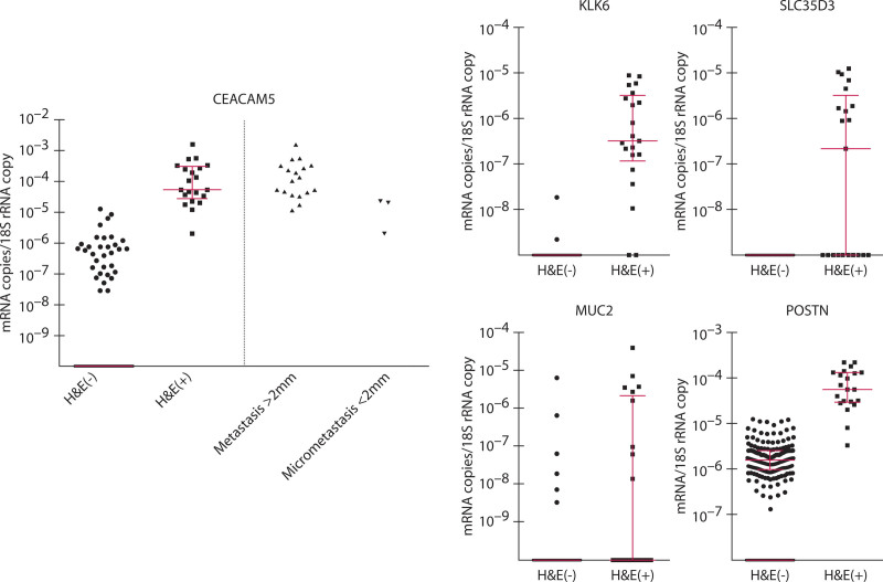
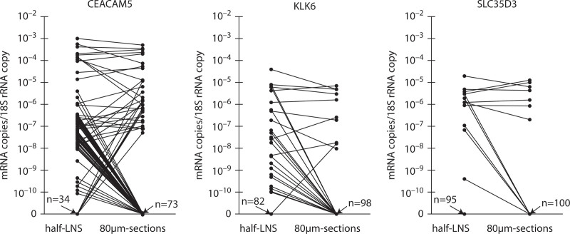
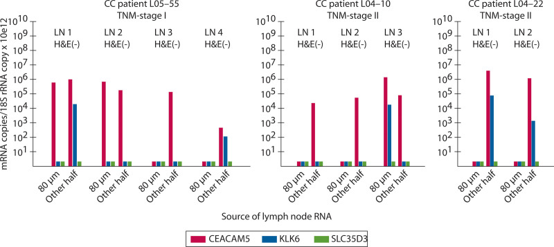
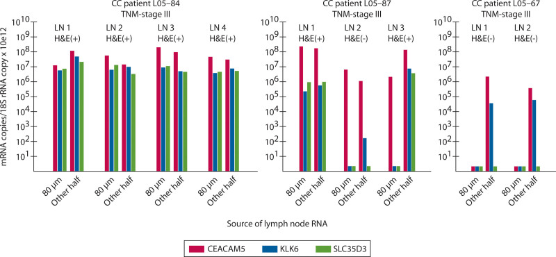
Results: The number of tumor cell-positive lymph nodes was 1.33-fold higher based on CEACAM5 mRNA levels compared with histopathological examination. Increasing the tissue volume analyzed for CEACAM5 levels from an 80-µm section to half a lymph node increased the number of positive nodes from 34 of 107 to 80 of 107 (p < 0.0001). Similarly, the number of positive nodes for the aggressiveness marker KLK6 increased from 9 of 107 to 24 of 107.

Limitations: Only a limited number of individual lymph nodes per patient was available for analysis.

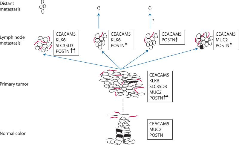
Conclusions: mRNA analysis of CEACAM5, KLK6, and SLC35D3 improves the detection of tumor cells in lymph nodes from patients surgically treated for colon cancer, and, together with POSTN and MUC2, it further allows characterization of the tumor cells with respect to aggressiveness and the tumor cell environment. See Video Abstract at http://links.lww.com/DCR/B650.

El anlisis de arnm de ceacam, klk, slcd, postn y muc mejora la deteccin y permite la caracterizacin de clulas tumorales en los ganglios linfticos de pacientes con cncer de colon: ANTECEDENTES:Las metástasis en los ganglios linfáticos son el factor de riesgo pronóstico más importante de recurrencia en pacientes con cáncer de colon que se han sometido a cirugía curativa. El método de rutina para detectar células tumorales diseminadas en los ganglios linfáticos es el examen microscópico de una o algunas secciones de tejido teñidas con hematoxilina-eosina por un patólogo capacitado. Sin embargo, este método es insensible principalmente porque se examina menos del 1% del volumen de los ganglios linfáticos, lo que conduce a una clasificación errónea.OBJETIVO:Investigar si el análisis de un grupo seleccionado de ARNm de biomarcadores mejora la detección y caracterización de metástasis / micrometástasis en los ganglios linfáticos en comparación con el método de rutina.DISEÑO:Una comparación en paralelo del análisis de ARNm de biomarcadores y la histopatología de 185 ganglios linfáticos de pacientes con cáncer de colon que representan las etapas I-IV, e investigación de la importancia del volumen de tejido de los ganglios linfáticos para la detección de células tumorales.ENTORNO CLINICO:Estudio colaborativo entre un hospital central de alto volumen y una institución universitaria preclínica.PACIENTES:Cincuenta y siete pacientes que han sido sometidos a resección tumoral por cáncer de colon.PRINCIPALES MEDIDAS DE VALORACION:copias de ARNm / copia de ARNr 18S de CEACAM5, KLK6, SLC35D3, POSTN y MUC2 mediante análisis múltiple y metástasis / micrometástasis detectadas por histopatología.RESULTADOS:El número de ganglios linfáticos con células tumorales positivas fue 1,33 veces mayor según los niveles de ARNm de CEACAM5 en comparación con el examen histopatológico. El aumento del volumen de tejido analizado para los niveles de CEACAM5 de una sección de 80 µm a la mitad de un ganglio linfático aumentó el número de ganglios positivos de 34/107 a 80/107 (p <0,0001). De manera similar, el número de nodos positivos para el marcador de agresividad KLK6 aumentó de 9/107 a 24/107.LIMITACIONES:Solo un número limitado de ganglios linfáticos individuales / paciente estuvo disponible para el análisis.CONCLUSIONES:El análisis de ARNm de CEACAM5, KLK6 y SLC35D3 mejora la detección de células tumorales en los ganglios linfáticos de pacientes con cáncer de colon tratados quirúrgicamente y, junto con POSTN y MUC2, permite además la caracterización de las células tumorales con respecto a la agresividad y el entorno celular tumoral. Consulte Video Resumen en http://links.lww.com/DCR/B650.


Figures

FIGURE 1.
FIGURE 1.
Study design and description of the clinical material. H&E = hematoxylin-eosin; OD260 = optical density at 260 nm; qRT-PCR = quantitative reverse transcription polymerase chain reaction.
FIGURE 2.
FIGURE 2.
All lymph nodes positive for disseminated tumor cells by microscopic examination of H&E-stained sections had high CEACAM5 mRNA levels. The first of 9 consecutive sections was stained by H&E and examined microscopically for the presence (H&E(+); n = 21) or absence (H&E(–); n = 164) of tumor cells. RNA extracted from the next 8 sections (80-µm section) were analyzed for amounts of CEACAM5, KLK6, SLC35D3, POSTN, and MUC2 mRNAs and 18S rRNA by multiplex qRT-PCR. Expression levels are given as biomarker mRNA copies per 18S rRNA copy. Long, red horizontal bar represents median and the short red horizontal bars represent the 25th and 75th percentile of levels of the indicated biomarker mRNA in H&E(+) and H&E(–) LNs. Each dot represents 1 LN. H&E = hematoxylin-eosin; LN = lymph node; qRT-PCR = quantitative reverse transcription polymerase chain reaction.
FIGURE 3.
FIGURE 3.
Many CEACAM5-, KLK6-, and SLC35D3-positive lymph nodes are missed in RNA extracted from the small volume of 8 lymph node tissue sections (80-µm sections) compared with RNA extracted from half the lymph node. RNA was extracted from one-half (half-LNs) of 107 LNs and from 8 consecutive, 10-µm-thick sections from the other half of the LN (80-µm sections). Levels, expressed as biomarker mRNA copies per 18S rRNA copy, of CEACAM5, KLK6, and SLC35D3 mRNAs were determined by multiplex qRT-PCR. Each dot represents 1 LN. Lines connect the mRNA levels in half-LN RNA extracts and the corresponding 80-µm section RNA extract of the other half of the LN. n values with arrows pointing to the x axis give the number of LNs with no detectable mRNA for the indicated biomarker in half-LN and 80-µm section RNA extracts. LN = lymph node; qRT-PCR = quantitative reverse transcription polymerase chain reaction.
FIGURE 4.
FIGURE 4.
Detection of lymph nodes of patients with stage I and II CC harboring aggressive tumor cells, as indicated by KLK6 mRNA, is increased when RNA is extracted from half a lymph node compared with a few tissue sections. Expression levels of CEACAM5 (red bars), KLK6 (blue bars), and SLC35D3 (green bars) mRNA in 2 to 4 LNs (LN1–LN4) of 1 patient with TNM stage I CC and 2 patients with stage II. Each LN was evaluated for the presence (H&E(+)) or absence (H&E(–)) of disseminated tumor cells by microscopic examination of H&E-stained tissue sections and analyzed for mRNA expression levels in RNA extracted from 80-µm sections adjacent to the section for H&E examination (80 µm) and in RNA extracted from the entire other half of the LN (Other half). Amounts of mRNA and 18S rRNA were determined by multiplex qRT-PCR and results are given as (mRNA copies/18S rRNA copy) × 1012. Short bars, below the 10 tick indicate no detection of the mRNA. CC = colon cancer; H&E = hematoxylin-eosin; LN = lymph node; qRT-PCR = quantitative reverse transcription polymerase chain reaction.
FIGURE 5.
FIGURE 5.
Detection of H&E(–) lymph nodes of patients with stage III CC harboring aggressive tumor cells, as indicated by KLK6 mRNA, is increased when RNA is extracted from half a lymph node compared with a few tissue sections. Expression levels of CEACAM5 (red bars), KLK6 (blue bars), and SLC35D3 (green bars) mRNA in 2 to 4 LNs (LN1–LN4) of 3 patients with stage III disease. Each LN was evaluated for the presence (H&E(+)) or absence (H&E(–)) of disseminated tumor cells by microscopic examination of H&E-stained tissue sections and analyzed for mRNA expression levels in RNA extracted from 80-µm sections adjacent to the section for H&E examination (80 µm) and in RNA extracted from the entire other half of the LN (Other half). Amounts of mRNA and 18S rRNA were determined by multiplex qRT-PCR and results are given as (mRNA copies/18S rRNA copy) × 1012. Short bars, below the 10 tick indicate no detection of the mRNA. CC = colon cancer; H&E = hematoxylin-eosin; LN = lymph node; qRT-PCR = quantitative reverse transcription polymerase chain reaction.
FIGURE 6.
FIGURE 6.
Expression of CEACAM5, KLK6, SLC35D3, POSTN, and MUC2 mRNAs at different stages of tumor development and metastasis in colon cancer: a hypothetical scenario. Normal colon: The epithelial cells all express CEACAM5 at the plasma membrane and several of them have differentiated to goblet cells producing mucin-2 (MUC2), the major mucin of colon. Only a few fibroblasts express periostin (POSTN), a ligand for αVβ3 and αVβ5 integrins, to support adhesion and migration of epithelial cells. Primary tumor: Epithelial cells that have transformed to cancer cells retain their expression of CEACAM5 and, in varying numbers, also produce MUC2. Fibroblasts in the microenvironment of the tumor cells strongly increase their expression of POSTN, thereby supporting tumor growth. In the growing tumor there is occasional induction of kallikrein-related peptidase 6 (KLK6) expression, that by its proteolytic activity facilitates escape of tumor cells to the draining lymphatics. Expression of solute carrier family 35 member D3 (SLC35D3), an orphan nucleotide sugar transporter with unknown function in the tumor cells is sporadically induced in KLK6-expressing cells. Lymph node metastasis: Tumor cells arriving to the lymph node induce elevated POSTN expression in resident fibroblasts, which in turn supports the establishment of micrometastases/metastases, and POSTN expression is increased further with the growing metastasis in a vicious circle. Tumor cells expressing KLK6 are prone to escape also from the metastasis spreading through lymphatics and blood to distant sites where metastases develop. It is possible that tumor cells not expressing KLK6 also leave the lymph node metastasis although likely at a lower frequency. Tumor cells expressing MUC2 are likely to be stationary, with a low degree of proliferation and a high degree of differentiation devoted to mucin production. Open and filled circles = tumor cells and normal epithelial cells; filled circles = MUC2-expressing cells; red elongated structures = POSTN expressing fibroblasts; fat black arrows = increased POSTN expression level; blue arrows = dissemination of tumor cells by lymphatics; hatched line = transformation of colonic epithelial cells to primary colon cancer tumor cells through several steps.

References

    1. Chang GJ, Rodriguez-Bigas MA, Skibber JM, Moyer VA. Lymph node evaluation and survival after curative resection of colon cancer: systematic review. J Natl Cancer Inst. 2007;99:433–441. - PubMed
    1. Iddings D, Bilchik A. The biologic significance of micrometastatic disease and sentinel lymph node technology on colorectal cancer. J Surg Oncol. 2007;96:671–677. - PubMed
    1. Nicastri DG, Doucette JT, Godfrey TE, Hughes SJ. Is occult lymph node disease in colorectal cancer patients clinically significant? A review of the relevant literature. J Mol Diagn. 2007;9:563–571. - PMC - PubMed
    1. Jemal A, Siegel R, Ward E, Murray T, Xu J, Thun MJ. Cancer statistics, 2007. CA Cancer J Clin. 2007;57:43–66. - PubMed
    1. Böckelman C, Engelmann BE, Kaprio T, Hansen TF, Glimelius B. Risk of recurrence in patients with colon cancer stage II and III: a systematic review and meta-analysis of recent literature. Acta Oncol. 2015;54:5–16. - PubMed

MeSH terms