. 2021 Aug;52(8):2637-2648.
doi: 10.1161/STROKEAHA.121.034362. Epub 2021 Jul 1.

Aflibercept, a VEGF (Vascular Endothelial Growth Factor)-Trap, Reduces Vascular Permeability and Stroke-Induced Brain Swelling in Obese Mice

Affiliations

Aflibercept, a VEGF (Vascular Endothelial Growth Factor)-Trap, Reduces Vascular Permeability and Stroke-Induced Brain Swelling in Obese Mice

Il-Doo Kim et al. Stroke. 2021 Aug.

Abstract

Background and purpose: Brain edema is an important underlying pathology in acute stroke, especially when comorbidities are present. VEGF (Vascular endothelial growth factor) signaling is implicated in edema. This study investigated whether obesity impacts VEGF signaling and brain edema, as well as whether VEGF inhibition alters stroke outcome in obese subjects.

Methods: High-fat diet-induced obese mice were subjected to a transient middle cerebral artery occlusion. VEGF-A and VEGFR2 (receptor) expression, infarct volume, and swelling were measured 3 days post-middle cerebral artery occlusion. To validate the effect of an anti-VEGF strategy, we used aflibercept, a fusion protein that has a VEGF-binding domain and acts as a decoy receptor, in human umbilical vein endothelial cells stimulated with rVEGF (recombinant VEGF; 50 ng/mL) for permeability and tube formation. In vivo, aflibercept (10 mg/kg) or IgG control was administered in obese mice 3 hours after transient 30 minutes middle cerebral artery occlusion. Blood-brain barrier integrity was assessed by IgG staining and dextran extravasation in the postischemic brain. A separate cohort of nonobese (lean) mice was subjected to 40 minutes middle cerebral artery occlusion to test the effect of aflibercept on malignant infarction.

Results: Compared with lean mice, obese mice had increased mortality, infarct volume, swelling, and blood-brain barrier disruption. These outcomes were also associated with increased VEGF-A and VEGFR2 expression. Aflibercept reduced VEGF-A-stimulated permeability and tube formation in human umbilical vein endothelial cells. Compared with the IgG-treated controls, mice treated with aflibercept had reduced mortality rates (40% versus 17%), hemorrhagic transformation (43% versus 27%), and brain swelling (28% versus 18%), although the infarct size was similar. In nonobese mice with large stroke, aflibercept neither improved nor exacerbated stroke outcomes.

Conclusions: The study demonstrates that aflibercept selectively attenuates stroke-induced brain edema and vascular permeability in obese mice. These findings suggest the repurposing of aflibercept to reduce obesity-enhanced brain edema in acute stroke.

Keywords: brain edema; comorbidity; obesity; stroke; vascular endothelial growth factor.


Figures

Figure 1.
Figure 1.
Impact of obesity on stroke outcomes. A, Survival curve for 72 h following transient middle cerebral artery occlusion (MCAO) shows increased mortality in obese mice (18 died out of 40) compared with lean mice (4 died out of 24); asterisk indicates P<0.05. B, Obese mice had increased rates of hemorrhagic transformation (which included hemorrhagic infarction [light red] and parenchymal hematoma [dark red]; n=10 and 13 for lean and obese, respectively). Arrowheads indicate petechial bleeding, and the arrow identifies hematoma. C, Obese mice had larger average infarct volumes and swelling when compared with lean mice. These outcomes were not correlated in individual obese mice while they were in individual lean mice (n=10 and 13 for lean and obese mice, respectively). D, IgG immunostaining (upper) and fluorescein isothiocyanate (FITC)-labeled dextran extravasation (lower) reveal increased vascular permeability in the obese mice (n=6 and 8 for lean and obese, respectively). *, **P<0.05, 0.01 vs lean.
Figure 2.
Figure 2.
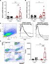
Effect of obesity on stroke-induced VEGF (vascular endothelial growth factor)-A and VEGFR2 (VEGF-A and receptor) expression in the brain. A and B, VEGF-A and VEGFR2 mRNA levels at 3 d post-middle cerebral artery occlusion (MCAO; n=7/group), respectively. C and D, Flow cytometry analysis of VEGFR2+ endothelial cells in the postischemic brain. C, Endothelial cells express CD31+ but do not express CD45. Scattered plot of isolated CD31+ cells from each hemisphere by CD31+ magnetic beads. Antibody controls to identify CD45 and VEGFR2 positive staining. D, Circles indicate CD45 and VEGFR2+ cells in CD31+ population. Flow cytometry and quantification of VEGFR2+ cells in CD31+/CD45 endothelial cell population. n=5/group. ctrl indicates contralateral; and ipsl, ipsilateral. *, **P<0.05, 0.01 vs lean, ##, ###P<0.01, 0.001 vs ctrl.
Figure 3.
Figure 3.
Effect of aflibercept on rVEGF (recombinant vascular endothelial growth factor)-induced permeability and tube formation in human umbilical vein endothelial cells (HUVECs). A, HUVECs were cultured confluent monolayer in inserts and stimulated with rVEGF for either 3 or 6 h before permeability of the monolayer was determined with fluorescein isothiocyanate (FITC)-labeled dextran (70 kDa). B, HUVECs were stimulated with rVEGF for 3 h either in the presence or absence of aflibercept (1 μg/mL). C, Images and quantification of tube formation. Quantification of tube formation HUVECs stimulated with rVEGF for 10 h in the presence of either low or high doses of aflibercept (1 or 10 μg/mL) showed that aflibercept dose-dependently impeded tube formation (n=3 for each condition). *, **, ***, **** indicates P<0.01, 0.05, 0.001, 0.0001 vs each control, respectively.
Figure 4.
Figure 4.
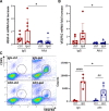
Effect of aflibercept on stroke-induced VEGF (vascular endothelial growth factor)-A and VEGFR2 (VEGF-A and receptor) expression in obese mice. A and B, Brain VEGF-A and VEGFR2 mRNA levels, respectively, in obese mice 3 d after middle cerebral artery occlusion (MCAO; n=7/group). C, Flow cytometry of CD31+ endothelial cells isolated from the poststroke brain using a CD31 magnetic beads. Circle indicates the number of VEGFR2+ cells in CD45/CD31+ endothelial cells. Note that the number of VGFR2+ cells in the ipsilateral (ipsl) hemisphere was significantly reduced in aflibercept-treated obese mice (n=5/group). ctrl indicates contralateral. *P<0.05 vs IgG, ##, #### P<0.01, 0.0001 vs ctrl.
Figure 5.
Figure 5.
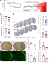
Effect of aflibercept on acute stroke outcomes in obese mice. A, Left, Survival curve for 72 h following transient middle cerebral artery occlusion (MCAO) in obese mice shows reduced mortality in aflibercept-treated mice (5/29 died) compared with control IgG-treated mice (12/27 died). Right, Incidence of hemorrhagic transformation at 3 d poststroke in survived animals. B, Neurological impairment score determined at 1 and 3 d after MCAO. C, Histological analyses for infarct volume and hemispheric brain swelling assessed 3 d after MCAO. D, Assessment of vascular permeability by IgG immunostaining (upper) and fluorescein isothiocyanate (FITC)-labeled dextran extravasation (lower; n=5–6./group). *, **P<0.05, 0.01, vs IgG.
Figure 6.
Figure 6.
Effect of aflibercept on acute stroke outcomes in nonobese mice with severe stroke. Mice subjected to 40 min middle cerebral artery occlusion (MCAO; severe stroke) were treated with IgG control or aflibercept and outcome analyses at 3 d poststroke. A, Assessment of infarct volume and percent hemispheric swelling. B, VEGFR2 (Vascular endothelial growth factor A and receptor) mRNA levels. C, Blood-brain barrier (BBB) permeability assessment. N=4–6/group. D, Survival curve. Eight out of 18 (IgG) and 4 out of 15 (aflibercept) were died for 3 d postischemia. *, ** P<0.05, 0.01 vs IgG.

References

    1. Paul S, Candelario-Jalil E. Emerging neuroprotective strategies for the treatment of ischemic stroke: an overview of clinical and preclinical studies. Exp Neurol. 2021; 335:113518. doi: 10.1016/j.expneurol.2020.113518 - PMC - PubMed
    1. Shi L, Rocha M, Leak RK, Zhao J, Bhatia TN, Mu H, Wei Z, Yu F, Weiner SL, Ma F, et al. . A new era for stroke therapy: integrating neurovascular protection with optimal reperfusion. J Cereb Blood Flow Metab. 2018; 38:2073–2091. doi: 10.1177/0271678X18798162 - PMC - PubMed
    1. O’Collins VE, Macleod MR, Donnan GA, Horky LL, van der Worp BH, Howells DW. 1,026 experimental treatments in acute stroke. Ann Neurol. 2006; 59:467–477. doi: 10.1002/ana.20741 - PubMed
    1. Pinto A, Tuttolomondo A, Di Raimondo D, Fernandez P, Licata G. Cerebrovascular risk factors and clinical classification of strokes. Semin Vasc Med. 2004; 4:287–303. doi: 10.1055/s-2004-861497 - PubMed
    1. Sarah K, McCann CBL. Comorbidity and age in the modelling of stroke: are we still failing to consider the characteristics of stroke patients? BMJ Open Sci. 2020; 4:e100013 - PMC - PubMed

Publication types

MeSH terms

Substances