. 2022 Feb;56(1):69-82.
doi: 10.1177/00236772211016922. Epub 2021 Jul 1.

Screening and validation of genome-edited animals

Affiliations

Screening and validation of genome-edited animals

Rosie K Bunton-Stasyshyn et al. Lab Anim. 2022 Feb.

Abstract

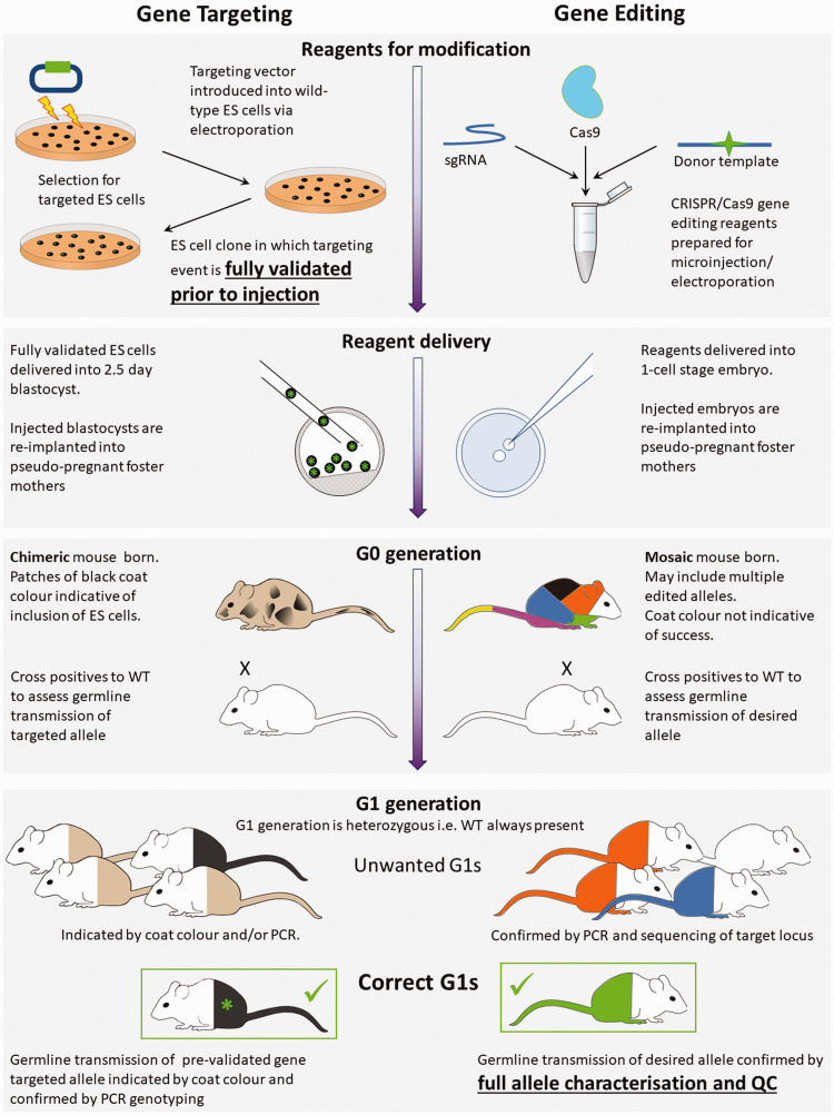
The emergence of an array of genome-editing tools in recent years has facilitated the introduction of genetic modifications directly into the embryo, increasing the ease, efficiency and catalogue of alleles accessible to researchers across a range of species. Bypassing the requirement for a selection cassette and resulting in a broad range of outcomes besides the desired allele, genome editing has altered the allele validation process both temporally and technically. Whereas traditional gene targeting relies upon selection and allows allele validation at the embryonic stem cell modification stage, screening for the presence of the intended allele now occurs in the (frequently mosaic) founder animals. Final confirmation of the edited allele can only take place at the subsequent G1 generation and the validation strategy must differentiate the desired allele from a range of unintended outcomes. Here we present some of the challenges posed by gene editing, strategies for validation and considerations for animal colony management.

L'émergence ces dernières années d'une gamme d'outils d'édition du génome a facilité l'introduction de modifications génétiques directement dans l'embryon, augmentant ainsi la facilité, l'efficacité et le catalogue des allèles accessibles aux chercheurs dans une gamme d'espèces. En contournant l'exigence d'une cassette de sélection et en permettant une large gamme de résultats outre l'allèle désiré, l'édition du génome a modifié le processus de validation d'allèle à la fois temporellement et techniquement. Alors que le ciblage traditionnel des gènes repose sur la sélection et permet une validation d'allèle au stade de la modification des cellules souches embryonnaires, le dépistage de la présence de l'allèle prévu se produit maintenant chez les animaux fondateurs (qui sont souvent mosaïques). La confirmation finale de l'allèle modifié ne peut avoir lieu qu'à la génération suivante (G1) et la stratégie de validation doit différencier l'allèle désiré d'une gamme de résultats inattendus. Nous présentons ici quelques-uns des défis posés par l'édition génétique, les stratégies de validation et les considérations pour la gestion des colonies animales.

Die Entwicklung einer Reihe von Tools zur Genom-Editierung hat in den letzten Jahren die Einführung von genetischen Modifikationen direkt in den Embryo begünstigt und die Einfachheit, Effizienz und den Katalog der Allele, die den Forschern über verschiedene Arten zugänglich sind, erhöht. Das Genome Editing umgeht die Notwendigkeit einer Selektionskassette und führt zu einer breiten Palette von Ergebnissen neben dem gewünschten Allel. Dadurch hat sich der Prozess der Allel-Validierung sowohl zeitlich als auch technisch verändert. Während das traditionelle Gen-Targeting auf Selektion beruht und eine Allel-Validierung im Stadium der ES-Zell-Modifikation ermöglicht, erfolgt das Screening auf das Vorhandensein des gewünschten Allels nun in den (häufig mosaischen) Gründertieren. Die endgültige Bestätigung des editierten Allels kann erst in der nachfolgenden G1-Generation erfolgen, und die Validierungsstrategie muss das gewünschte Allel von einer Reihe von unbeabsichtigten Ergebnissen unterscheiden. Hier stellen wir einige der Herausforderungen im Zusammenhang mit Gen-Editierung, Strategien zur Validierung und Überlegungen zum Management von Tierkolonien vor.

La emergencia en los últimos años de una serie de herramientas de modificación del genoma ha facilitado la introducción de modificaciones genéticas directamente en el embrión, aumentando así la facilidad, la eficiencia y las opciones de alelos accesibles para los investigadores en una gran variedad de especies. Al poder saltarnos la necesidad de un grupo de selección cerrado y con la posibilidad de una amplia gama de resultados además del alelo deseado, la modificación del genoma ha alterado el proceso de validación de alelos tanto temporal como técnicamente. Mientras que la selección tradicional de genes depende de la selección y permite la validación de alelos en la fase de modificación de células madre, hacer un cribado para identificar la presencia del alelo deseado ahora ocurre con los animales fundadores (normalmente mosaico). La confirmación final del alelo modificado solo puede hacerse en la siguiente generación G1 y la estrategia de validación debe diferenciar el alelo requerido de una serie de resultados no intencionados. En este estudio presentamos algunos de los retos que supone la edición genética, así como estrategias de validación y consideraciones para la gestión de colonias de animales.

Keywords: Animal model; GM; PCR; Quality assurance / control.


Conflict of interest statement

Declaration of Conflicting Interests: The author(s) declared no potential conflicts of interest with respect to the research, authorship, and/or publication of this article.

Figures

Figure 1.
Figure 1.
Gene targeting and genome editing processes. Comparison between traditional gene targeting and more recent gene-editing processes from reagents to correct G1 animals. The figure highlights the validation of embryonic stem (ES) cells prior to delivery into the embryo, whereas gene editing technologies rely on validation at the mouse stage. Left: in traditional gene targeting, validation occurs in embryonic stem (ES) cells, prior to their delivery into 2.5-day blastocysts. G0 animals are chimeric, being composed of two pre-defined cell types, those of the host and the validated ES cell. At the G1 generation there are only two possible genotypes to identify. ES cell and host with different coat colours can be used so that coat colour indicates incorporation of the ES cells. At G0 coat colour will be mixed at different ratios depending upon inclusion of ES cells into the embryo. At G1 full coat colour can demonstrate that ES cells have populated the germ-cells of the G0 parent and PCR genotyping can confirm which of the two ES cell derived alleles has been transmitted. Right: in gene editing, genome modification happens in vivo after reagents are delivered to the 1-cell stage embryo. G0 mice are mosaic, being composed of cells with multiple different genotypes. Multiple editing events during early embryonic development may produce an assortment of cell-lineages all with differing, and previously undefined, genotypes. At the G1 generation offspring with many different genotypes may be born and it is only at this stage that the desired allele can be definitively identified, and the mutation and background be fully validated. Coat colour cannot be used to indicate success of gene editing as there is no host-donor chimera. PCR, polymerase chain reaction; WT, wild type.
Figure 2.
Figure 2.
Editing strategies and assays for allele validation. Deletion: nucleases target either side of an exon or other region to be deleted. Polymerase chain reaction (PCR) primers flank targeted region and can detect a reduction in amplicon size after deletion. Sequence of the PCR amplicon should be confirmed via Sanger sequencing (or similar). Droplet digital (dd)PCR copy counting of the wild-type (WT) allele (blue assay) in G1s will identify a copy number of one. Point mutations and indels: a nuclease is targeted to a single location where the nucleotide change is to be made. PCR primers flank this location. A size shift will not be present in the desired mutant. The nucleotide change must be identified by Sanger sequencing of the amplicon. ddPCR copy counting of the WT allele (blue assay) in G1s will identify a copy number of one. When a repair donor is used to produce a specific mutation, ddPCR copy counting of the mutant sequence (orange) in G1s should give copy number of one. Large knock-ins: a nuclease is targeted to a single site for insertion of the knock-in. PCR primers flanking the target location (dark green) can be used to detect a size increase in the presence of an insertion. Primers specific to the repair template (light green) can detect donor insertion. Primer pairs combining one primer binding within the repair donor and another binding outside of the repair donor (light and dark green pairs) can identify on-target donor insertion. Sanger sequencing of these amplicons must be used to confirm identity and may require multiple Sanger reads depending on the insertion size. Long-read sequencing (purple) can identify the entire segment in a single read and confirm whether a fully correct allele is present in the G0 generation. ddPCR copy counting of both the WT allele (blue assay) and the repair donor (orange) in G1s should each give copy number of one. Floxed: nucleases target either side of an exon (or other region) where LoxP sites are to be inserted. PCR primers flanking the entire region (dark green) will amplify a larger product, but the ability to discriminate a size shift via standard agarose gel electrophoresis will depend on the relative size of the floxed region. A primer pair specific to the two LoxP insertions (light green) can identify insertion of a single long donor template, or in the case of two short donors, in cis insertion of both donors. Primer pairs combining one primer specific to a LoxP insertion (light green) and another within the flanking target locus (dark green) can identify on target integration. Sanger sequencing of these amplicons must be used to confirm identity and may require multiple Sanger reads depending on the insertion size. Long-read sequencing (purple) can identify the entire segment in a single read and confirm whether a fully correct allele is present in the G0 generation. ddPCR copy counting of the WT allele (blue assay) in G1s will identify a copy number of two. Assays specific to each LoxP insertion (orange and brown) will each give copy number of one. UTR, untranslated region.
Figure 3.
Figure 3.
Unintended mutations and methods to detect them. Different types of unintended mutation can occur depending on the editing strategy employed – the number of nuclease target cut sites and whether a repair template is included – while other mutations, such as large deletions, can occur in all cases. Here we present some common examples along with assays which can be used for their detection. Most simply, polymerase chain reaction (PCR) amplification of the targeted locus may identify unwanted insertions or deletion by a shift in band size. Sequencing PCR amplicons can reveal unintended indels and incorrect donor insertions. An inability to amplify an expected product (indicated by a red dotted line) may indicate a rearranged donor insertion (causing incompatible primer orientation) or a partial integration (failure to insert the primer binding region). Copy counting assays, using droplet digital (dd)PCR (or qPCR) are useful for identifying insertion events which are not readily detected by regular PCR. For example, a repair donor or a deleted region can reinsert randomly elsewhere in the genome and ddPCR assays can help to detect this. Concatemerized on target insertion of a donor can be challenging to identify by regular PCR due to amplification bias, while deletions which expand beyond the primers binding sites will be missed entirely. Both are readily detected by ddPCR.
Figure 4.
Figure 4.
Animals with visual phenotypes. A variety of phenotypes were observed in CRISPR G0 mice from four different projects. (a) Short faces, domed head and missing teeth were observed in 11 out of 24 animals born from a cytoplasmic CRISPR–Cas9 injection to introduce a point mutation into Csf1r. (b) Pups with hair loss and/or tufty hair across the whole body were seen in three out of 15 animals derived from a CRISPR–Cas9 pronuclear injection to introduce a point mutation in Foxn1. (c) Pup with abnormal hind legs and gait derived from CRISPR–Cas9 electroporation to introduce a point mutation into Itpr1. Hind feet point upwards and pup weight bears on hind heels, displaying abnormal movements and hopping. (d) Oedema and odd body shape were observed in all pups born from a cytoplasmic CRISPR–Cas9 injection to introduce a point mutation into Lemd2. Upon dissection, the liver was found to be enlarged.

References

    1. Robertson E, Bradley A, Kuehn M, et al. Germ-line transmission of genes introduced into cultured pluripotential cells by retroviral vector. Nature 1986; 323: 445–448. - PubMed
    1. Doetschman T, Gregg RG, Maeda N, et al. Targeted correction of a mutant HPRT gene in mouse embryonic stem cells. Nature 1987; 330: 576–578. - PubMed
    1. Valenzuela DM, Murphy AJ, Frendewey D, et al. High-throughput engineering of the mouse genome coupled with high-resolution expression analysis. Nat Biotechnol 2003; 21: 652–659. - PubMed
    1. Skarnes WC, et al. A conditional knockout resource for the genome-wide study of mouse gene function. Nature 2011; 474: 337–342. - PMC - PubMed
    1. Geurts AM, Rosen B, West AP, et al. Knockout rats via embryo microinjection of zinc-finger nucleases. Science 2009; 325: 433. - PMC - PubMed