Review
. 2021 Jun 30;19(1):67.
doi: 10.1186/s12964-021-00750-x.

Src family kinases, adaptor proteins and the actin cytoskeleton in epithelial-to-mesenchymal transition

Affiliations
Review

Src family kinases, adaptor proteins and the actin cytoskeleton in epithelial-to-mesenchymal transition

Maria A Ortiz et al. Cell Commun Signal. .

Abstract

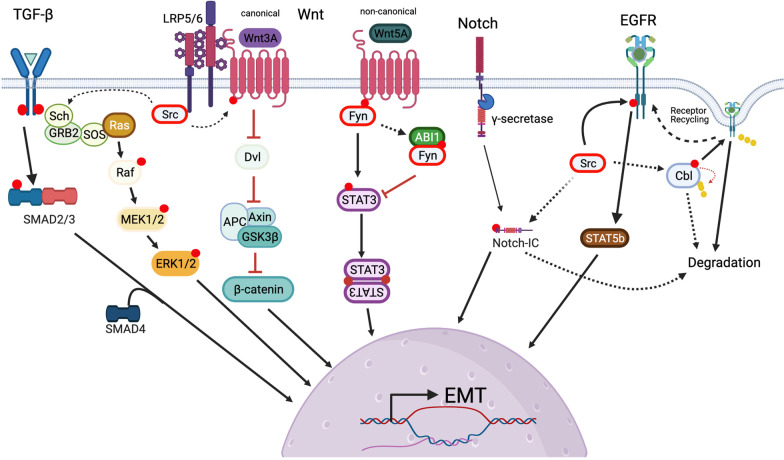
Over a century of scientific inquiry since the discovery of v-SRC but still no final judgement on SRC function. However, a significant body of work has defined Src family kinases as key players in tumor progression, invasion and metastasis in human cancer. With the ever-growing evidence supporting the role of epithelial-mesenchymal transition (EMT) in invasion and metastasis, so does our understanding of the role SFKs play in mediating these processes. Here we describe some key mechanisms through which Src family kinases play critical role in epithelial homeostasis and how their function is essential for the propagation of invasive signals. Video abstract.

Keywords: Actin cytoskeleton; Epithelial-to-mesenchymal transition; Invasion; Metastasis; Src family kinases; Treatment resistance; Unique domain.


Conflict of interest statement

The authors declare that they have no competing interests.

Figures

Fig. 1
Fig. 1
Primary structure of SRC family kinases. Domain and signal conservation within Src family kinases. Src family kinases are activated at the membrane, which involves lipid/myristate modification within the SH4 region and membrane binding. There is very little known about the UD, which might be also involved in membrane localization, activation and ligand substrate binding (see lower panel for LCK). SH3 and SH2 domain bind substrates and regulate the catalytic activity of the tyrosine kinase (domain). Posttranslational modifications in Src family kinases essential for membrane localization (myristylation and palmytoilation); activation (within kinase domain) and inhibition (C-terminal tail) (phosphorylation) are highlighted in the diagram. Domain/regulatory regions are depicted as lines and boxes: Src homology 1, SH1, tyrosine kinase/catalytic domain; SRC homology 2 or SH2; SRC homology 3, SH3; SRC homology 4, SH4, and unstructured Unique Domain, UD). Bottom panels: NMR Structural Ensemble of the C-terminal tail of (A) CD4 (red) or (B) CD8α (magenta) in complex with the intrinsically disordered Unique Domain of LCK (residues 7–35). Both complexes are very dynamic and are mediated by Zn (blue spheres). The pdb codes are 1Q68 and 1Q69 for CD4 and CD8α, respectively
Fig. 2
Fig. 2
Conformational changes associated with Src family kinase activation and inhibition. Left, inactive conformation of Src family kinases is associated with lack of membrane binding, lack of phosphorylation of the activation loop tyrosine, phosphorylation of the C-terminal regulatory region tyrosine; and characterized by a “closed conformation”. The closed conformation is maintained through inhibitory SH3-SH2 domain interactions with the catalytic domain (SH1), and the C-terminal regulatory phosphotyrosine interaction with the SH2 domain. These interactions prevent ligand substrate binding. Right, Active “open” conformation allows for ligand binding and autophosphorylation of the activation loop tyrosine. The balance of active/inactive conformation is regulated by Csk that promotes phosphorylation of C-terminal tyrosine; and its dephosphorylation by PTP1B/Shp1/2. Ligand binding facilitates activation but may also regulate kinase activity through restricting access (competitive inhibition) to the active catalytic domain. Examples of SRC kinase ligands/substrates are listed on the far right. Created with BioRender.com
Fig. 3
Fig. 3
Differential expression of Src family kinases at the tissue and cellular levels. Left panel, Protein Atlas aggregate data on expression pattern of Src family kinases in tissues. Based on these information four kinases, SRC, FYN, YES and LYN, can be considered ubiquitously expressed. While FGR, LCK, HCK, and LYN are more restricted to tissue associated with immune response and blood cell production (bone marrow and spleen), and lungs. Right panel, RNA-based single cell type expression data of SRC family kinases. These data also support general expression patterns of SFKs. Human Protein Atlas available from http://www.proteinatlas.org (Tissue atlas “protein expression overview” and cell type atlas “single cell types”; Gene entries: SRC, FYN, YES1, FGR, LYN, LCK, HCK, BLK)
Fig. 4
Fig. 4
Cell structures critical for epithelial homeostasis and regulated by the interface of SFKs and actin cytoskeleton regulatory complexes. Loss of integrity the structures and /or deregulation of these complexes promote EMT. Top left, Major cell adhesion and invasive phenotypes mediated by SFKs critical for the epithelial to mesenchymal transition. Top right, Cell–cell adhesion, with adherens junctions regulated by E-cadherin, catenins, p120. WAVE complex regulates actin cytoskeleton input into adherence junctions with ABI1 being a key adaptor protein for SRC at the membrane (see text for more details). Lower left, Active SRC regulates FAK to maintain and regulate integrin function. Integrins are intimately involved in cell migration, invasive potential and tissue specificity during metastasis. Lower right, a key complex that regulates invadopodia through SRC-Arg and SRC-Tsk5 axes: N-WASp, Arp2/3, cortactin and control actin polymerization input in invadopodia. MMP2 and MMP9 degrade collagen and other ECM integral proteins. Created with BioRender.com
Fig. 5
Fig. 5
Cell signals that regulate SFKs input into major cellular pathways. Roles of SFKs in modulating some of the key EMT-inducing pathways. Top, Signals initiated by a variety of receptors, TGF-beta, WNT pathway receptors (LRP5/6), Notch and growth factor receptors such as EGFR. All signals involve cytoplasmic regulators and modulators which end up activating transcription factors/coactivators to regulate gene expression. See text for details. Created with BioRender.com

References

    1. Rous P. A sarcoma of the fowl transmissible by an agent separable from the tumor cells. J Exp Med. 1911;13(4):397–411. doi: 10.1084/jem.13.4.397. - DOI - PMC - PubMed
    1. Oppermann H, Levinson AD, Varmus HE, Levintow L, Bishop JM. Uninfected vertebrate cells contain a protein that is closely related to the product of the avian sarcoma virus transforming gene (src) Proc Natl Acad Sci USA. 1979;76(4):1804–1808. doi: 10.1073/pnas.76.4.1804. - DOI - PMC - PubMed
    1. Talamonti MS, Roh MS, Curley SA, Gallick GE. Increase in activity and level of pp60c-src in progressive stages of human colorectal cancer. J Clin Invest. 1993;91(1):53–60. doi: 10.1172/JCI116200. - DOI - PMC - PubMed
    1. Thiery JP, Acloque H, Huang RY, Nieto MA. Epithelial-mesenchymal transitions in development and disease. Cell. 2009;139(5):871–890. doi: 10.1016/j.cell.2009.11.007. - DOI - PubMed
    1. Saitoh M. Involvement of partial EMT in cancer progression. J Biochem. 2018;164(4):257–264. doi: 10.1093/jb/mvy047. - DOI - PubMed

Publication types

MeSH terms

Substances

LinkOut - more resources