

Cardiovascular magnetic resonance imaging in children after recovery from symptomatic COVID-19 or MIS-C: a prospective study
- PMID: 34193197
- PMCID: PMC8245157
- DOI: 10.1186/s12968-021-00786-5
Cardiovascular magnetic resonance imaging in children after recovery from symptomatic COVID-19 or MIS-C: a prospective study
Abstract
Background: Cardiac evaluations, including cardiovascular magnetic resonance (CMR) imaging and biomarker results, are needed in children during mid-term recovery after infection with SARS-CoV-2. The incidence of CMR abnormalities 1-3 months after recovery is over 50% in older adults and has ranged between 1 and 15% in college athletes. Abnormal cardiac biomarkers are common in adults, even during recovery.
Methods: We performed CMR imaging in a prospectively-recruited pediatric cohort recovered from COVID-19 and multisystem inflammatory syndrome in children (MIS-C). We obtained CMR data and serum biomarkers. We compared these results to age-matched control patients, imaged prior to the SARS-CoV-2 pandemic.
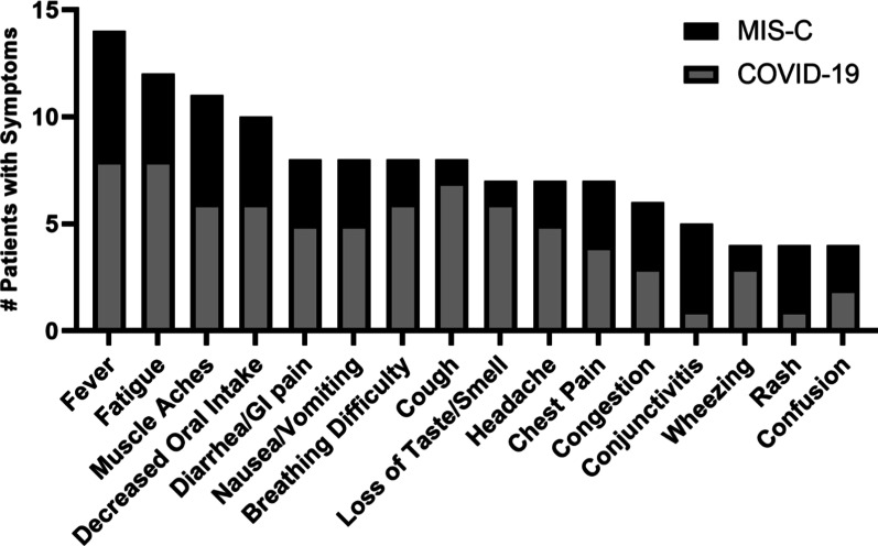
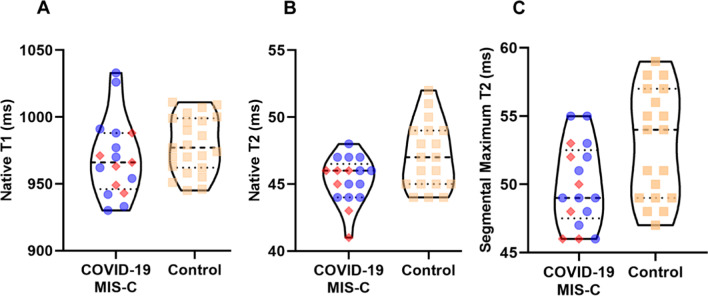
Results: CMR was performed in 17 children (13.9 years, all ≤ 18 years) and 29 age-matched control patients without SARS-CoV-2 infection. Cases were recruited with symptomatic COVID-19 (11/17, 65%) or MIS-C (6/17, 35%) and studied an average of 2 months after diagnosis. All COVID-19 patients had been symptomatic with fever (73%), vomiting/diarrhea (64%), or breathing difficulty (55%) during infection. Left ventricular and right ventricular ejection fractions were indistinguishable between cases and controls (p = 0.66 and 0.70, respectively). Mean native global T1, global T2 values and segmental T2 maximum values were also not statistically different from control patients (p ≥ 0.06 for each). NT-proBNP and troponin levels were normal in all children.
Conclusions: Children prospectively recruited following SARS-CoV-2 infection had normal CMR and cardiac biomarker evaluations during mid-term recovery. Trial Registration Not applicable.
Keywords: COVID-19; Cardiac magnetic resonance imaging; MIS-C; Pediatric; SARS-CoV-2.
Conflict of interest statement
The authors declare that they have no competing interests.
Figures
References
Publication types
MeSH terms
Substances
Supplementary concepts
Grants and funding
LinkOut - more resources
Full Text Sources
Medical
Research Materials
Miscellaneous
