. 2021 Aug 25;41(34):7314-7325.
doi: 10.1523/JNEUROSCI.0684-21.2021. Epub 2021 Jun 30.

Corticotropin-Releasing Hormone from the Pontine Micturition Center Plays an Inhibitory Role in Micturition

Affiliations

Corticotropin-Releasing Hormone from the Pontine Micturition Center Plays an Inhibitory Role in Micturition

Jason P Van Batavia et al. J Neurosci. .

Abstract

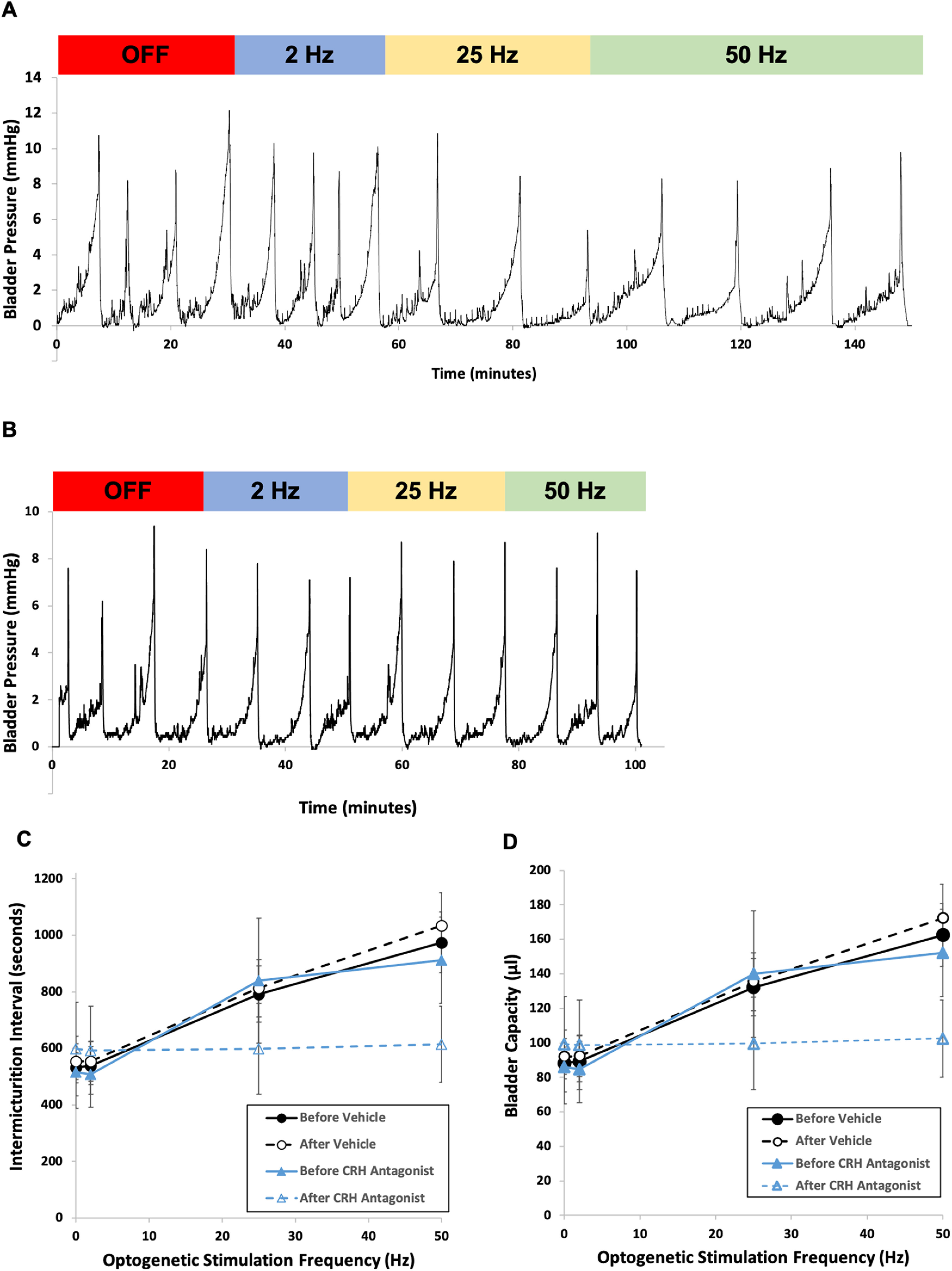
Lower urinary tract or voiding disorders are prevalent across all ages and affect >40% of adults over 40 years old, leading to decreased quality of life and high health care costs. The pontine micturition center (PMC; i.e., Barrington's nucleus) contains a large population of neurons that localize the stress-related neuropeptide, corticotropin-releasing hormone (CRH) and project to neurons in the spinal cord to regulate micturition. How the PMC and CRH-expressing neurons in the PMC control volitional micturition is of critical importance for human voiding disorders. To investigate the specific role of CRH in the PMC, neurons in the PMC-expressing CRH were optogenetically activated during in vivo cystometry in unanesthetized mice of either sex. Optogenetic activation of CRH-PMC neurons led to increased intermicturition interval and voided volume, similar to the altered voiding phenotype produced by social stress. Female mice showed a significantly more pronounced phenotype change compared with male mice. These effects were eliminated by CRH-receptor 1 antagonist pretreatment. Optogenetic inhibition of CRH-PMC neurons led to an altered voiding phenotype characterized by more frequent voids and smaller voided volumes. Last, in a cyclophosphamide cystitis model of bladder overactivity, optogenetic activation of CRH-PMC neurons returned the voiding pattern to normal. Collectively, our findings demonstrate that CRH from PMC spinal-projecting neurons has an inhibitory function on micturition and is a potential therapeutic target for human disease states, such as voiding postponement, urinary retention, and underactive or overactive bladder.SIGNIFICANCE STATEMENT The pontine micturition center (PMC), which is a major regulator of volitional micturition, is neurochemically heterogeneous, and excitatory neurotransmission derived from PMC neurons is thought to mediate the micturition reflex. In the present study, using optogenetic manipulation of CRH-containing neurons in double-transgenic mice, we demonstrate that CRH, which is prominent in PMC-spinal projections, has an inhibitory function on volitional micturition. Moreover, engaging this inhibitory function of CRH can ameliorate bladder hyperexcitability induced by cyclophosphamide in a model of cystitis. The data underscore CRH as a novel target for the treatment of voiding dysfunctions, which are highly prevalent disease processes in children and adults.

Keywords: Barrington's nucleus; lower urinary tract symptoms; micturition; neuro-urology; optogenetics; pontine micturition center.


Figures

Figure 1.
Figure 1.
Cre-lox recombination results in ChR2 expression in the majority of CRH-expressing neurons in the PMC. A, Schematic of coronal section through mouse brain at the level of the pons illustrating the location of the pontine micturition center (green) and the adjacent locus ceruleus (red). B, Using RNAscope to perform ISH, probes targeting the mRNA for CRH identify PMC (green) and probes targeting the mRNA for TH identifies the LC (red). C, In situ hybridization (ISH) of CRH mRNA (red) and ChR2 (green) shows significant overlap of neurons with both colors in PMC (200×). Scale bar, 50 µm. First panel shows DAPI (blue) only, second panel CRH (red) only, third panel ChR2 (green) only, and fourth panel overlap of all 3 channels.
Figure 2.
Figure 2.
Effect of optogenetic stimulation of the PMC on urodynamics. A, B, Representative cystometry tracings of bladder pressure versus time in a control male mouse (ChR2 only) (A) and a double-transgenic CRH-ChR2 male mouse (B). Baseline cystometry pattern is obtained at an infusion rate of 10 µl/min with the optogenetic probe turned off. After establishment of baseline IMI and bladder capacity, the LED generator is turned on and delivers light via the fiberoptic probe to PMC at the frequencies denoted above the traces. A, For the control male mouse, the IMI remained constant with the optogenetic stimulation off and at all frequencies of stimulation. B, For the CRH-ChR2 male mouse, there was an increase in both IMI and bladder capacity with 25 Hz (yellow shaded area of graph) or 50 Hz (green shaded area of graph) stimulation. C, Box-and-whisker plot showing the mean effects of optogenetic photostimulation on IMI in control and CRH-ChR2 mice. For control mice (ChR2 only, n = 6, 3 male, 3 female), there was no effect of frequency on IMI (F(3,3) = 2.1, p = 0.28). In contrast, for double CRH-ChR2 mice (n = 15, 8 male, 7 female), there was an effect of Hz (F(3,12) = 48, p < 0.001). A two-way repeated-measures ANOVA revealed an effect of genotype (F(1,19) = 5.7, p = 0.027) and genotype × Hz interaction (F(3,17) = 21, p < 0.0001). There were statistically significant differences in IMI between baseline and optogenetic stimulation at both 25 and 50 Hz (p < 0.0001; Mann–Whitney U test). D, Similarly, box-and-whisker plot shown for bladder capacity. There was no effect of Hz for control mice (F(3,3) = 2.1, p = 0.28). In contrast, there was an effect of Hz for CRH-ChR2 mice (F(3,12) = 47, p < 0.0001). A two-way repeated-measures ANOVA revealed an effect of genotype (F(1,19) = 5.7, p = 0.028) and genotype × Hz interaction (F(3,17) = 20, p < 0.0001). CRH-ChR2 mice had significantly larger bladder capacities at both 25 Hz and 50 Hz optogenetic stimulation compared with baseline (p < 0.0001; Mann–Whitney U test). *** denotes that the difference between control and CRH-ChR2 values at that Hz was significant (Mann-Whitney U test, p < 0.001).
Figure 3.
Figure 3.
Fluoroscopy during cystometry study showed complete bladder emptying in mice with each voiding cycle. The male CRH-ChR2 mouse was placed in a Plexiglas cage atop the X-ray fluoroscopy detector, and cystometry studies were repeated while infusing a radiopaque contrast agent. Single or spot X-ray images were obtained at various points during the filling and voiding cycle at baseline and at each optogenetic stimulation frequency. Voids were detected by spikes in vesical pressure on the cystometry curve as well as by visually detecting voids on paper placed on the bottom of the cage. A, In this fluoroscopic image, the mouse has a total bladder volume of 120 μl (red arrow). B, Upon voiding, the bladder was completely empty with no residual contrast seen on repeat fluoroscopic image (red arrow indicates location of bladder, now empty). Also seen in these views are the fiberoptic probe (yellow arrows) leading to the mouse's head as well as an anchoring bone screw placed to help the dental cement hold the cannula in place.
Figure 4.
Figure 4.
Effects of CRH antagonist on urodynamics during optogenetic stimulation of the pontine micturition center of a double-transgenic CRH-ChR2 female mouse. A, Graph showing cystometry tracing in a double-transgenic CRH-ChR2 female mouse. Baseline urodynamics were established with the optogenetic probe turned off and then at increasing stimulation frequencies as denoted above the tracing. IMI increased at both 25 and 50 Hz stimulation. B, On the following day, CRH antagonist NBI-30775 was administered (10 mg/kg in 10% solution of 0.1 m tartaric acid in sterile water, i.p.) to the same female mouse from A. One hour later, baseline urodynamics were established with the optogenetic probe turned off and then with increasing stimulation frequencies. In the presence of the CRH antagonist, no changes in IMI were noted at any optogenetic stimulation frequency. C, Graph showing the effects of optogenetic stimulation on IMI in double-transgenic CRH-ChR2 mice before and after vehicle control injection (n = 4, 2 male, 2 female; solid black and dashed black lines, respectively) and before and after CRH antagonist injection (n = 4, 2 male, 2 female; solid blue and dashed blue lines, respectively). A two-way repeated-measures ANOVA revealed an effect of Hz (F(3,4) = 55, p = 0.001) but not before/after vehicle control condition × Hz interaction was seen (F(3,4) = 0.4, p = 0.77). For mice before CRH antagonist, there was an effect of HZ on IMI (F(3,4) = 173, p < 0.01). A two-way repeated-measures ANOVA revealed a before/after CRH antagonist condition × Hz interaction (F(3,4) = 121, p < 0.01). D, Graph showing the effects of optogenetic stimulation on bladder capacity in CRH-ChR2 mice before and after vehicle control (n = 4; solid black and dashed black lines, respectively) and before and after CRH antagonist injection (n = 4; solid blue and dashed blue lines, respectively). A two-way repeated-measures ANOVA revealed an effect of Hz (F(3,4) = 53, p = 0.001) on bladder capacity but not before/after vehicle control condition × Hz interaction was seen (F(3,4) = 0.47, p = 0.7). For mice before CRH antagonist, there was an effect of Hz on bladder capacity (F(3,4) = 181, p < 0.01). A two-way repeated-measures ANOVA revealed a before/after CRH antagonist condition × Hz interaction (F(3,4) = 146, p < 0.01). Points in graphs represent mean values. Error bars indicate ± SEM.
Figure 5.
Figure 5.
Effect of optogenetic inhibition of the PMC on urodynamics. A, B, Representative cystometry tracings of bladder pressure versus time in a control male mouse (ArchT only) (A) and a double-transgenic CRH-ArchT male mouse (B). Baseline cystometry pattern is obtained at an infusion rate of 10 µl/min with the optogenetic probe turned off. After establishment of baseline IMI and bladder capacity, the LED generator (530 nm) is turned on and delivers light via the optic fiber to PMC at the frequencies denoted above the traces. A, For the control male mouse, the IMI remained constant with the optogenetic stimulation off and at all frequencies of stimulation. B, For the CRH-ArchT male mouse, there was a decrease in both IMI and bladder capacity with 25 Hz (yellow shaded area of graph) or 50 Hz (green shaded area of graph) stimulation. As mentioned in Materials and Methods, only photostimulation off and at 50 Hz was used in the final analysis for simplicity given equivalence of off with 2 and 25 Hz with 50 Hz. C, Box-and-whisker plot showing effects of optogenetic stimulation on IMI in control and CRH-ArchT mice. For control mice (CRH-Cre only, n = 6, 3 male, 3 female), there was no effect of photostimulation status on IMI (F(1,3) = 0.3, p = 0.99). In contrast, for double CRH-ArchT mice (n = 8, 4 male, 4 female), there was an effect of photostimulation status (F(1,5) = 5.1, p = 0.006). A two-way repeated-measures ANOVA revealed an effect of genotype (F(1,12) = 23, p < 0.001). In CRH-ArchT mice, there were statistically significant differences in IMI between baseline and optogenetic photostimulation at 50 Hz (p < 0.01; Mann–Whitney U test). D, Box-and-whisker plot for bladder capacity based on mouse type and photostimulation status. There was no effect of photostimulation status for control mice (F(1,3) = 0.3, p = 0.99). In contrast, there was an effect of photostimulation status for CRH-ArchT mice (F(1,5) = 4.3, p < 0.01). A two-way repeated-measures ANOVA revealed an effect of genotype (F(1,12) = 21, p < 0.001). CRH-ArchT mice had significantly larger bladder capacities of 50 Hz photostimulation compared with baseline (p < 0.001; Mann–Whitney U test). *** denotes that the difference in IMI (part C) and bladder capacity (part D) between control and CRH-ArchT mice was significant at 50Hz (Mann-Whitney U test, p < 0.001 for both).
Figure 6.
Figure 6.
Optogenetic stimulation of CRH neurons in the PMC reverses the voiding phenotype of cyclophosphamide-induced cystitis. The 24 h voiding micturograms obtained using UroVoid (Med Associates) metabolic chambers from the same 8-week-old CRH-Ch2R male mouse at (A) baseline, (B) 48 h after i.p. cyclophosphamide instillation, and (C) after start of optogenetic photostimulation at 50 Hz. Graphs showing (D) voiding frequency per 24 h period, (E) mean voided volume, and (F) mean IMI at baseline, 48 h after intraperitoneal CPM administration, and during optogenetic photostimulation in CRH-Ch2R mice (n = 6, 3 male, 3 female).

Similar articles

Cited by

References

    1. Asok A, Draper A, Hoffman AF, Schulkin J, Lupica CR, Rosen JB (2018) Optogenetic silencing of a corticotropin-releasing factor pathway from the central amygdala to the bed nucleus of the stria terminalis disrupts sustained fear. Mol Psychiatry 23:914–922. 10.1038/mp.2017.79 - DOI - PMC - PubMed
    1. Bangasser DA, Curtis A, Reyes BA, Bethea TT, Parastatidis I, Ischiropoulos H, Van Bockstaele EJ, Valentino RJ (2010) Sex differences in corticotropin-releasing factor receptor signaling and trafficking: potential role in female vulnerability to stress-related psychopathology. Mol Psychiatry 15:896–904. 10.1038/mp.2010.66 - DOI - PMC - PubMed
    1. Bangasser DA, Reyes BA, Piel D, Garachh V, Zhang XY, Plona ZM, Van Bockstaele EJ, Beck SG, Valentino RJ (2013) Increased vulnerability of the brain norepinephrine system of females to corticotropin-releasing factor overexpression. Mol Psychiatry 18:166–173. 10.1038/mp.2012.24 - DOI - PMC - PubMed
    1. Barrington FJ (1925) The effect of lesions of the hind- and mid-brain on micturition in the cat. Exp Physiol 15:81–102. 10.1113/expphysiol.1925.sp000345 - DOI
    1. Bjorling DE, Wang ZY, Bushman W (2011) Models of inflammation of the lower urinary tract. Neurourol Urodyn 30:673–682. 10.1002/nau.21078 - DOI - PMC - PubMed

Publication types

MeSH terms