Prediction of successful caudal epidural injection using color Doppler ultrasonography in the paramedian sagittal oblique view of the lumbosacral spine
- PMID: 34193640
- PMCID: PMC8255148
- DOI: 10.3344/kjp.2021.34.3.339
Prediction of successful caudal epidural injection using color Doppler ultrasonography in the paramedian sagittal oblique view of the lumbosacral spine
Abstract
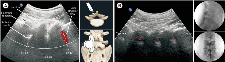
Background: Ultrasound-guided caudal epidural injection (CEI) is limited in that it cannot confirm drug distribution at the target site without fluoroscopy. We hypothesized that visualization of solution flow through the inter-laminar space of the lumbosacral spine using color Doppler ultrasound alone would allow for confirmation of drug distribution. Therefore, we aimed to prospectively evaluate the usefulness of this method by comparing the color Doppler image in the paramedian sagittal oblique view of the lumbosacral spine (LS-PSOV) with the distribution of the contrast medium observed during fluoroscopy.
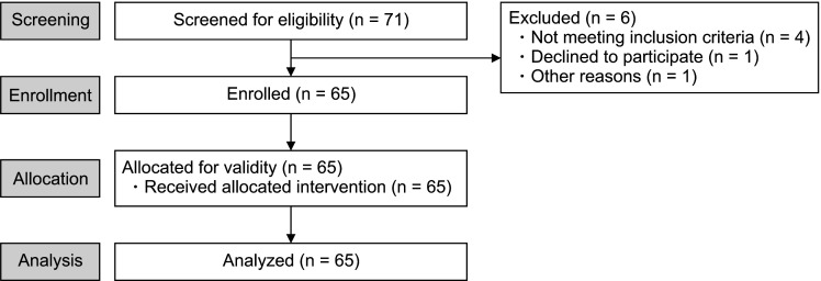
Methods: Sixty-five patients received a 10-mL CEI of solution containing contrast medium under ultrasound guidance. During injection, flow was observed in the LSPSOV using color Doppler ultrasonography, following which it was confirmed using fluoroscopy. The presence of contrast image at L5-S1 on fluoroscopy was defined as "successful CEI." We then calculated prediction accuracy for successful CEI using color Doppler ultrasonography in the LS-PSOV. We also investigated the correlation between the distribution levels measured via color Doppler and fluoroscopy.
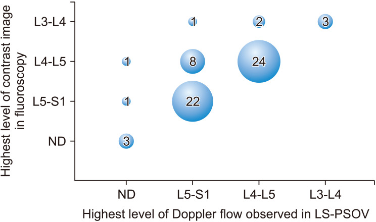
Results: Prediction accuracy with color Doppler ultrasonography was 96.9%. The sensitivity, specificity, positive predictive value, and negative predictive value were 96.7%, 100%, 100%, and 60.0%, respectively. In 52 of 65 patients (80%), the highest level at which contrast image was observed was the same for both color Doppler ultrasonography and fluoroscopy.
Conclusions: Our findings demonstrate that color Doppler ultrasonography in the LS-PSOV is a new method for determining whether a drug solution reaches the lumbosacral region (i.e. , the main target level) without the need for fluoroscopy.
Keywords: Anesthesia; Caudal; Color; Contrast Media; Doppler; Epidural; Equivalence Trial; Fluoroscopy; Injections; Interventional.; Low Back Pain; Lumbar Vertebrae; Sensitivity and Specificity; Ultrasonography.
Conflict of interest statement
No potential conflict of interest relevant to this article was reported.
Figures
Similar articles
-
The Role of Power Doppler Ultrasonography in Caudal Epidural Injection.Medicina (Kaunas). 2022 Apr 22;58(5):575. doi: 10.3390/medicina58050575. Medicina (Kaunas). 2022. PMID: 35629992 Free PMC article.
-
The feasibility of color Doppler ultrasonography for caudal epidural steroid injection.Pain. 2005 Nov;118(1-2):210-4. doi: 10.1016/j.pain.2005.08.014. Epub 2005 Oct 4. Pain. 2005. PMID: 16213088
-
Predicting Epidural Space Spread Using Ultrasound Color Doppler Imaging in Interlaminar Epidural Steroid Injection: A Prospective Observational Study.Pain Physician. 2022 Mar;25(2):E349-E356. Pain Physician. 2022. PMID: 35322990
-
Image Guidance Technologies for Interventional Pain Procedures: Ultrasound, Fluoroscopy, and CT.Curr Pain Headache Rep. 2018 Jan 26;22(1):6. doi: 10.1007/s11916-018-0660-1. Curr Pain Headache Rep. 2018. PMID: 29374352 Review.
-
Ultrasound- versus fluoroscopy-guided injections in the lower back for the management of pain: a systematic review.Eur Radiol. 2019 Jul;29(7):3401-3409. doi: 10.1007/s00330-019-06065-3. Epub 2019 Mar 18. Eur Radiol. 2019. PMID: 30887198
Cited by
-
The value of the peroneus brevis tendon cross-sectional area in early diagnosing of peroneus brevis tendinitis: The peroneus brevis tendon cross-sectional area.Medicine (Baltimore). 2022 Oct 28;101(43):e31276. doi: 10.1097/MD.0000000000031276. Medicine (Baltimore). 2022. PMID: 36316917 Free PMC article.
-
Ultrasound-guided caudal epidural injection to treat symptoms of lumbar spinal stenosis: a retrospective study.Eur J Transl Myol. 2024 May 7;34(2):12167. doi: 10.4081/ejtm.2024.12167. Eur J Transl Myol. 2024. PMID: 38713057 Free PMC article.
-
Comparison of international medical costs for interventional pain treatment: a focus on Korea and Japan.Korean J Pain. 2024 Jan 1;37(1):51-58. doi: 10.3344/kjp.23254. Epub 2023 Dec 11. Korean J Pain. 2024. PMID: 38072796 Free PMC article.
-
The Power of Color Flow Doppler Ultrasonography Versus Blind Technique in Localization of Epidural Catheter: A Randomized Prospective Study.Anesth Pain Med. 2024 Jun 30;14(3):e147828. doi: 10.5812/aapm-147828. eCollection 2024 Jun. Anesth Pain Med. 2024. PMID: 39416806 Free PMC article.
References
LinkOut - more resources
Full Text Sources