. 2021 Jun 30;12(1):4047.
doi: 10.1038/s41467-021-24292-5.

Recapitulation of selective nuclear import and export with a perfectly repeated 12mer GLFG peptide

Affiliations

Recapitulation of selective nuclear import and export with a perfectly repeated 12mer GLFG peptide

Sheung Chun Ng et al. Nat Commun. .

Abstract

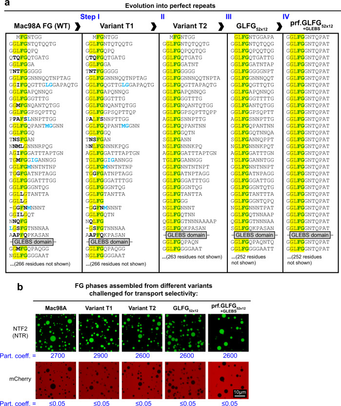
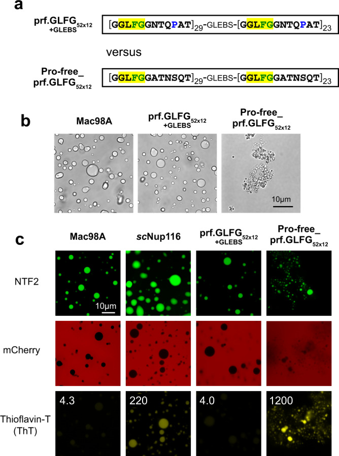
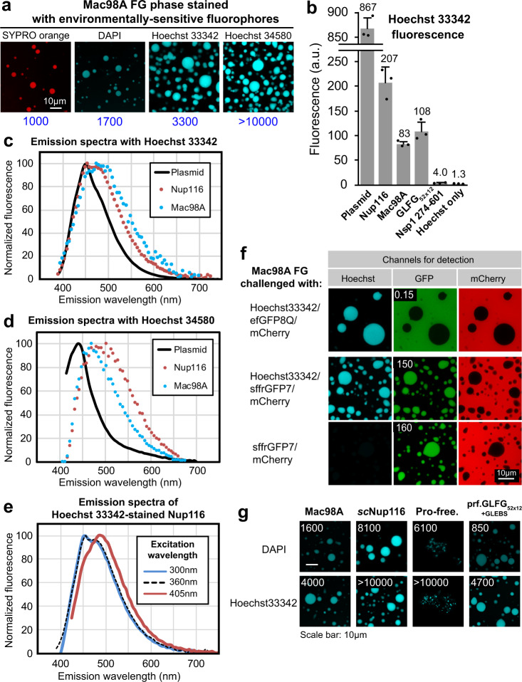
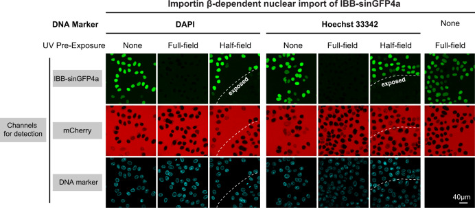
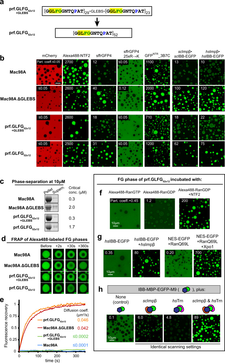
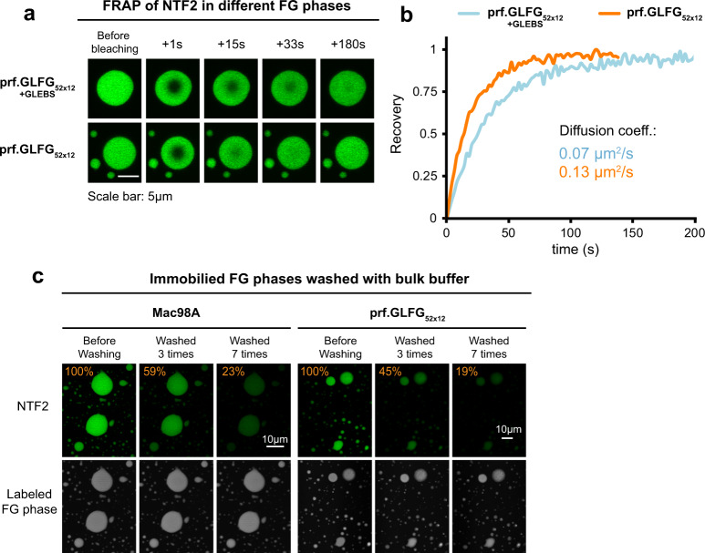
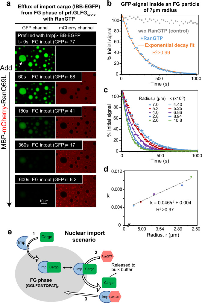
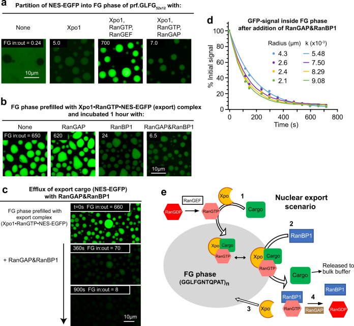
The permeability barrier of nuclear pore complexes (NPCs) controls nucleocytoplasmic transport. It retains inert macromolecules while allowing facilitated passage of importins and exportins, which in turn shuttle cargo into or out of cell nuclei. The barrier can be described as a condensed phase assembled from cohesive FG repeat domains. NPCs contain several distinct FG domains, each comprising variable repeats. Nevertheless, we now found that sequence heterogeneity is no fundamental requirement for barrier function. Instead, we succeeded in engineering a perfectly repeated 12mer GLFG peptide that self-assembles into a barrier of exquisite transport selectivity and fast transport kinetics. This barrier recapitulates RanGTPase-controlled importin- and exportin-mediated cargo transport and thus represents an ultimately simplified experimental model system. An alternative proline-free sequence forms an amyloid FG phase. Finally, we discovered that FG phases stain bright with 'DNA-specific' DAPI/ Hoechst probes, and that such dyes allow for a photo-induced block of nuclear transport.


Conflict of interest statement

The authors declare no competing interests.

Figures

Fig. 1
Fig. 1. Engineering of a perfectly repeated GLFG domain with authentic phase behavior and NPC-typical selectivity.
a Sequences of the Tetrahymena thermophila MacNup98A (“Mac98A”) FG domain and simplified variants. Mac98A contains an intervening 44-residue GLEBS domain (binding site for the mRNA export mediator Gle2p) that was kept unchanged in these variants. For space economy, only the N-terminal ≈400 residues up to the GLEBS domain are shown. The C-terminal sequences of the FG domains (≈270 residues not shown) follow the same design principle (see Supplementary Note 1 for complete sequences and Supplementary Table 1 for amino acid compositions). b Indicated FG domains were dissolved at a concentration of 1 mM in 4 M guanidinium hydrochloride, and phase separation was initiated by a rapid 50-fold dilution with assay buffer (50 mM Tris/HCl pH 7.5 50 mM, 150 mM NaCl, 5 mM DTT), followed by another fourfold dilution in buffer +6 µM mCherry and 1 µM Alexa Fluor 488-labeled rat NTF2. Samples were analyzed by confocal laser-scanning microscopy (CLSM). All FG domain variants phase-separated to µm-sized, spherical FG particles that excluded mCherry (red) very well and accumulated the NTR NTF2 (green) to very high partition coefficients. Note that phase separation occurred here already at 5 µM FG domain concentration, which is at least 100 times lower than the most conservative estimate for the local Nup98 FG domain concentration at NPCs (48 copies in a cylinder of 70 nm diameter and 40 nm height).
Fig. 2
Fig. 2. Absence of prolines in a perfectly repetitive GLFG variant favors amyloid-like structures.
a Comparison of two perfectly repetitive FG domains. The “Pro-free_prf.GLFG52x12” is a proline-free variant (see also Supplementary Table 1). b Phase-contrast images of FG phases assembled from Mac98A FG domain, the proline-containing prf.GLFG52x12[+GLEBS] and the Pro-free_prf.GLFG52x12. Note that the former two formed spherical particles, but the Pro-free variant formed irregular shapes. The experiment was repeated independently four times with similar results, and representative images are shown. c FG phases were assembled from the indicated FG domains and challenged with Alexa488-labeled NTF2, mCherry, and Thioflavin-T (ThT). Numbers refer to the ratios of fluorescence inside the FG phases to that in the surrounding buffer. Images were taken at 30 min after FG phase assembly. Note that the bright ThT stain of the Pro-free variant is diagnostic of amyloid structures. The N/Q-rich Nup116 FG repeats served as a ThT positive control. See also Supplementary Figs. 1 and 2.
Fig. 3
Fig. 3. FG phases stained by fluorescent aromatic compounds.
a Mac98A FG particles were formed, incubated with 2 µM of indicated fluorophores, and analyzed by CLSM. Note that all fluorophores got attracted by the phase; however, there were great differences with Atto488 being nearly inert and Cy3 accumulating very strongly. Table 1 lists all fluorophores tested and their partition coefficients. Chemical structures are shown in Supplementary Fig. 3. * indicates that a cysteine-quenched fluorophore maleimide was tested. Blue numbers: ratios of signals inside Mac98A FG phase to signal in the surrounding buffer. Scan settings/image brightness were adjusted individually. b scNup116 FG particles were challenged with efGFP8Q, or variants with an added C-terminal cysteine that had been modified either with maleimidopropionic acid (“Mal-quenched”), Atto488-, Alexa647-, Atto647N-, and Cy3-maleimides, respectively. Blue numbers: ratios of GFP fluorescence inside: outside the Nup116 FG phase. The far-red channel detects signals from Alexa647/Atto647N (colored cyan), and the red channel detects signals from Cy3 (colored magenta). Scanning settings/image brightness were adjusted individually. Note that, e.g., the Atto647N modification increased the GFP partition coefficient ≈30-fold.
Fig. 4
Fig. 4. Bright staining of condensed FG phases with “DNA-specific” DAPI and Hoechst dyes.
a Environmentally sensitive fluorophores (SYPRO orange, DAPI, and Hoechst dyes) were used to stain Mac98A FG particles. Note the extremely high fluorescence in:out ratios (blue numbers). b Bulk fluorescence of FG domains (2.67 mg/ml) stained with 20 µM Hoechst 33342. Excitation was at 360 nm, detection at 440–480 nm. Data are presented as mean values (arbitrary unit, a.u.) of three independent replicates, with individual data points shown as dots. Error bars are the standard deviations. Note that the cohesive FG domains (Nup116, Mac98A, and GLFG52x12) gave strong signals, while the signal for non-cohesive Nsp1 FG fragment (residue 274–601) was at least 20 times weaker. Source data are provided as a Source Data file. c Emission spectra of Hoechst 33342-stained plasmid, Nup116 and Mac98A FG phases (excitation wavelength: 360 nm). Note the red-shifted fluorescence of the FG phases. d Emission spectra of Hoechst 34580-stained plasmid, Nup116 and Mac98A FG phases (excitation wavelength: 405 nm). e Emission spectra of Hoechst 33342-stained Nup116 FG phase recorded at three distinct excitation wavelengths: 360 nm corresponds to the absorption peak (see Supplementary Fig. 4) and excitation with a shorter wavelength (300 nm) showed essentially the same emission spectrum. However, excitation with a longer wavelength (405 nm, at “red edge” of the excitation spectrum) led to a red-shift of the emission spectrum and of the emission peak, known as the “red edge effect”. Source data (ce) are provided as a Source Data file. f Mac98A FG phase was challenged with the indicated probes and imaged by confocal microscopy. sffrGFP7 is an FG-philic GFP variant. Detection wavelengths: Hoechst/410–470 nm, GFP/500–555 nm, and mCherry/577–700 nm. Note that the presence of Hoechst did not bias the partitioning of either sffrGFP7 or mCherry. g Different FG phases were stained with DAPI/Hoechst. Image brightness was adjusted individually. “Pro-free.”: Pro-free_prf.GLFG52x12. Numbers refer to FG in:out fluorescence ratios.
Fig. 5
Fig. 5. UV-induced blocking of NPCs by DAPI/ Hoechst.
HeLa cells were grown in cell culture wells, permeabilized with digitonin, and preincubated without or with the traditional DNA markers DAPI or Hoechst 33342. Indicated wells were then partially exposed for 30 s with UV light from the microscope’s mercury/metal halide lamp (wavelength ≈ 365 nm). A mixture of importin β, IBB-sinGFP4a (an importin β-dependent import cargo), mCherry (as a passive exclusion marker), components of the Ran system, and an ATP/GTP-regenerating system (“Methods”) was added. Import, followed live by confocal laser-scanning microscopy, was allowed to proceed at 21 °C. The micrographs show the ≈200 s time point of either a non-UV exposed area or a fully exposed one (“full-field”). “Half-field” indicates the region where the illumination boundary (dashed line) had crossed. Strong accumulation of IBB-sinGFP4a was observed in nuclei not pre-exposed to UV or samples without DAPI/Hoechst addition. The combination of DAPI or Hoechst with UV illumination blocked the import of IBB-sinGFP4a completely.
Fig. 6
Fig. 6. Selectivity of FG phases assembled from Pro-containing perfect repeats.
a An ultimately simplified FG domain (prf.GLFG52x12) was generated by deleting the GLEBS domain from prf.GLFG52x12[+GLEBS]. b FG phases were challenged with different protein probes for selectivity. Scanning settings/image brightness were adjusted individually due to the large range of signals. Note that GLEBS-free FG phases (Mac98A ΔGLEBS and prf.GLFG52x12) allowed for a stronger accumulation of NTRs, NTR·cargo complexes, and the NTR-like sffrGFP4. However, the conversion of the Mac98A FG domain to perfect repeats caused only small changes in NTR-accumulation. Scale bar: 10 μm. sc: Saccharomyces cerevisiae; hs: Homo sapiens. c In total, 10 µM of indicated FG domain variants were allowed to phase separate and the FG phases were pelleted by ultracentrifugation. Equivalent ratios of pellets and supernatants were analyzed by SDS-PAGE/Coomassie-staining. Critical concentrations for phase separation were taken as the concentrations that remained in the supernatants (“Methods”). The experiment was repeated independently three times with similar results, and representative images are shown. Full scans of gels with molecular weight markers were provided as a Source Data file. d FG particles spiked with 2% Alexa488-(covalently) labeled FG domains (of the same species) were photobleached. Fluorescence recovery after photobleaching (FRAP) was recorded over time. Scale bar: 3 μm. e Recovery curves corresponding to (d). Y axis: recovery was normalized to 1 for a complete recovery. The numbers are translational diffusion coefficients derived from fitting the datasets to theoretical recovery curves (“Methods”). The GLEBS-containing Mac98A and prf.GLFG52x12[+GLEBS] FG domains were essentially immobile. Source data are provided as a Source Data file. f Alexa488-labeled Ran was converted either to the RanGTP form (by pre-incubation with RanGEF and an ATP/GTP-regenerating system), or to the RanGDP form (by pre-incubation with RanGAP). Panels show the partitioning of these Ran forms into the ultimately simplified prf.GLFG52x12 phase. Where indicated, unlabeled NTF2 (2 µM dimer concentration) was also added. g prf.GLFG52x12 phase was challenged with import (IBB-EGFP) and export cargoes (EGFP fused to a strong Xpo1-dependent nuclear export signal (NES) + RanQ69L, a mutant of Ran locked in the GTP form) in the absence or presence of the corresponding NTRs: Impβ/Xpo1. h prf.GLFG52x12 phase was challenged with an 80 kDa import cargo carrying two orthogonal import signals, an IBB (recognized by Impβ) and an M9 domain (recognized by transportin, Trn). Note that strong intra-phase accumulation was only observed in the presence of both NTRs, recapitulating the requirement for large cargo transport through NPCs. Numbers refer to GFP fluorescence ratios in the central regions of the particles to that in the surrounding buffer.
Fig. 7
Fig. 7. Mobility of NTRs inside FG phases assembled from Pro-containing perfect repeats.
a, b FG particles (unlabeled) assembled from both prf.GLFG52x12 versions were preloaded with Alexa488-labeled NTF2, bleached, and FRAP was recorded over time. Experiments were repeated with multiple FG particles (n = 5). Representative images (a) and recovery curves (b) are shown. See Supplementary Figs. 5 and 6 for the complete dataset and other FRAP datasets for GFP-NTR derivatives. Source data are provided as a Source Data file. c FG particles spike with Alexa647 (covalently)-labeled FG domain molecules (FG-tracers) were immobilized on glass, preloaded with Alexa488-labeled NTF2, washed multiple times with fresh buffer (“Methods”) while the fluorescence signals were recorded. Orange numbers: % Alexa488 signal inside FG phase relative to the initial signal. For both Mac98A and prf.GLFG52x12 (GLEBS-free), signals of NTF2 within FG particles dropped to ≈20% of the original after seven rounds of washing. The majority of FG particles remained immobilized after washing, indicating their stickiness to the support.
Fig. 8
Fig. 8. Recapitulation of RanGTPase-controlled nuclear import with the FG phase assembled by the perfectly repetitive variant.
a The FG phase of prf.GLFG52x12 was initially loaded with hsImpβ·hsIBB-EGFP complex. At time = 0 s, a RanQ69L fragment fused to MBP-mCherry was added, which triggered the unloading of IBB-EGFP from Impβ and thus an efflux from the FG phase. Fluorescence signals of GFP and mCherry were recorded over time. The ratios of GFP fluorescence inside:outside an FG particle of 7 μm radius (marked with a white arrow) were quantified. Note that some Ran fusion was recruited to the FG phase by Impβ but only arrested at the rim of the FG particles, as expected from the FG-phobic effect of the MBP-mCherry group. This was meant to ensure that the RanGTP-reaction occurs only at the particles’ surface. b Time course of IBB-EGFP signal inside the FG phase. Blue colored: GFP signal inside an FG particle of 7 μm radius (marked with a white arrow in (a)), normalized to % of the initial signal. Gray: a control set without RanGTP addition. Orange: best-fit to a single exponential decay function: ft=Aekt+B, where t is the time, A = 110, B = 0, k = 4.4 × 10−3. c The GFP signals inside FG particles with different radii were fitted to the single exponential decay function to obtain the respective time constants k. Source data are provided as a Source Data file. d k obtained in the experiment described above was plotted against 1/r2, where r is the FG particle’s radius. e Illustration of the above processes.
Fig. 9
Fig. 9. Recapitulation of RanGTPase-controlled nuclear export with the FG phase formed by the perfectly repetitive variant.
a The FG phase of prf.GLFG52x12 was incubated for 60 min with NES-EGFP, an ATP/GTP-regenerating system, and either (i) buffer only, (ii) Xpo1, (iii) Xpo1 and wild-type RanGTP (preloaded with GTP by RanGEF), or (iv) Xpo1 and RanGDP (converted by RanGAP to the GDP form). b In another setup, the FG phase of prf.GLFG52x12 was initially filled with the Xpo1·RanGTP·NES-EGFP complex in the presence of RanGEF, then incubated individually for 60 min with the indicated components, and finally imaged. c, d As in (b), the FG phase of prf.GLFG52x12 was initially filled with the Xpo1·RanGTP·NES-EGFP complex. At time=0 s, RanGAP and RanBP1 were added to trigger NES-EGFP efflux. The signal of GFP was recorded over time and analyzed as described in Fig. 8. Source data are provided as a Source Data file. d A plot of GFP signals inside FG particles of different radii against time and best-fits of the single exponential decay function: ft=Aekt+B (see also Supplementary Fig. 9). e Illustration of the above processes.

References

    1. Feldherr CM. The nuclear annuli as pathways for nucleocytoplasmic exchanges. J. Cell Biol. 1962;14:65–72. doi: 10.1083/jcb.14.1.65. - DOI - PMC - PubMed
    1. Hoelz A, Glavy JS, Beck M. Toward the atomic structure of the nuclear pore complex: when top down meets bottom up. Nat. Struct. Mol. Biol. 2016;23:624–630. doi: 10.1038/nsmb.3244. - DOI - PMC - PubMed
    1. Hampoelz B, Andres-Pons A, Kastritis P, Beck M. Structure and assembly of the nuclear pore complex. Annu. Rev. Biophys. 2019;48:515–536. doi: 10.1146/annurev-biophys-052118-115308. - DOI - PubMed
    1. Lin DH, Hoelz A. The structure of the nuclear pore complex (an update) Annu. Rev. Biochem. 2019;88:725–783. doi: 10.1146/annurev-biochem-062917-011901. - DOI - PMC - PubMed
    1. Mohr D, Frey S, Fischer T, Güttler T, Görlich D. Characterisation of the passive permeability barrier of nuclear pore complexes. EMBO J. 2009;28:2541–2553. doi: 10.1038/emboj.2009.200. - DOI - PMC - PubMed

Publication types