. 2021 Jun 30;11(1):13586.
doi: 10.1038/s41598-021-93033-x.

Evaluation of different grafting materials for alveolar cleft repair in the context of orthodontic tooth movement in rats

Affiliations

Evaluation of different grafting materials for alveolar cleft repair in the context of orthodontic tooth movement in rats

Stephan Christian Möhlhenrich et al. Sci Rep. .

Abstract

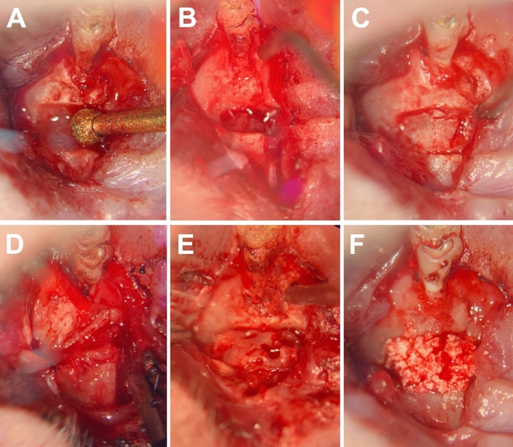
To minimize the postoperative risks posed by grafting autologous transplants for cleft repair, efforts are being made to improve grafting materials for use as potential alternatives. The aim of this study was to compare the bone graft quality of different bone substitutes including the gold standard autografts during the healing processes after cleft repair in the context of orthodontic treatment. In 21 Wistar rats, a complete, continuity-interrupting cleft was created. After 4 weeks, cleft repair was performed using autografts from the hips' ischial tuberosity, human xenografts, or synthetic bone substitutes [beta-tricalcium phosphate (β-TCP)/hydroxyapatite (HA)]. After another 4 weeks, the first molar movement was initiated in the reconstructed jaw for 8 weeks. The bone remodeling was analyzed in vivo using micro-computed tomography (bone mineral density and bone volume fraction) and histology (new bone formation). All the grafting materials were statistically different in bone morphology, which changed during the treatment period. The β-TCP/HA substitute demonstrated less resorption compared to the autologous and xenogeneic/human bone, and the autografts led to a stronger reaction in the surrounding bone. Histologically, the highest level of new bone formation was found in the human xenografts, and the lowest was found in the β-TCP/HA substitute. The differences between the two bone groups and the synthetic materials were statistically significant. Autografts were confirmed to be the gold standard in cleft repair with regard to graft integration. However, parts of the human xenograft seemed comparable to the autografts. Thus, this substitute could perhaps be used as an alternative after additional tissue-engineered modification.


Conflict of interest statement

The authors declare no competing interests.

Figures

Figure 1
Figure 1
Timeline of the animal cleft research procedure: the thick, solid lines represent the experimental measures under intraperitoneal injection, such as cleft creation, cleft repair, application of orthodontic appliances, killing, and resection, while the dotted lines represent the radiological follow-up monitoring in µ-CT under isoflurane anesthesia.
Figure 2
Figure 2
View of the operative situs of the left maxilla in supine position (magnification × 10): first molar above, mouth tip below: (A) artificial alveolar cleft creation using an ultrasonic device; (B) alveolar cleft with intact mucosa to the maxillary sinus and nasal passage; and (C) artificial alveolar cleft filled with bone wax. Re-entry and cleft repair were performed with (D) autograft from the ischial tuberosity of the hip, (E) human xenograft, or (F) β-TCP/HA bone substitute material.
Figure 3
Figure 3
(A) Applied orthodontic appliance based on a 0.14 N nickel–titanium closed coil tension spring fixed between the first molar and the incisors using tension springs after conditioning of the teeth through acid etching using 39% phosphonic acid and bonding agent and dental composite (magnification × 4). (B) Anterior moved first molar after 8 weeks of orthodontic tooth movement (magnification × 6).
Figure 4
Figure 4
Three-dimensional micro-CT volume rendering after (A) cleft creation (µCT T0) and immediately after cleft repair (µCT T1) using (B) autologous bone (green), (C) xenogeneic/human bone (red), and (D) synthetic (β-TCP/HA) bone substitute (blue) and the surrounding alveolar bone (beige area).
Figure 5
Figure 5
Sagittal view of the CT scans after cleft creation (µCT T0) and cleft repair with and without an orthodontic appliance (µCT T1, T3) for analyzing the bone quality of the augmented bone in the cleft (green area: autologous bone; red area: xenogeneic bone; blue area: synthetic bone substitute) and the surrounding alveolar bone (beige area) (magnification × 40).
Figure 6
Figure 6
Histological cross-section (toluidine blue stains) through the reconstructed jaw 84 days after cleft repair using autologous bone (A,D), xenogeneic/human bone (B,E), and synthetic tricalcium phosphate/hydroxyapatite bone substitute (C,F): overview: (AC) × 100 magnification; detailed view: (DF) up to × 350 magnification, persistent bone/substitute (*), new bone formation (arrows).
Figure 7
Figure 7
Radiological changes of the bone mineral density (BMD) and bone volume fraction (BV/TV) in the grafting materials (A,B) and in the bone surrounding the cleft (C,D) of the maxillary reconstruction: column bars of the mean values and p-values for the comparisons between the three different materials at seven points in the 84-day healing period after cleft repair.
Figure 8
Figure 8
Radiological bone changes in the (A) bone mineral density (BMD) and (B) bone volume fraction (BV/TV) of the grafting materials and the bone surrounding the cleft in the maxillary reconstruction: line diagram of the mean values in the context of the bone structural morphology behavior over time.
Figure 9
Figure 9
Results of the histological structural analysis of the reconstructed maxilla with regard to persistent grafting material and new bone formation: column bars of the mean values (A) or the corresponding percentage (B) and p-values for the comparisons of the three materials after the 84-day cleft repair healing period.

References

    1. Bajaj AK, Wongworawat AA, Punjabi A. Management of alveolar clefts. J. Craniofac. Surg. 2003;14:840–846. doi: 10.1097/00001665-200311000-00005. - DOI - PubMed
    1. Aalami OO, et al. Applications of a mouse model of calvarial healing: Differences in regenerative abilities of juveniles and adults. Plast. Reconstr. Surg. 2004;114:713–720. doi: 10.1097/01.prs.0000131016.12754.30. - DOI - PubMed
    1. Sharif F, Ur Rehman I, Muhammad N, MacNeil S. Dental materials for cleft palate repair. Mater. Sci. Eng. C Mater. Biol. Appl. 2016;61:1018–1028. doi: 10.1016/j.msec.2015.12.019. - DOI - PubMed
    1. Canady JW, Zeitler DP, Thompson SA, Nicholas CD. Suitability of the iliac crest as a site for harvest of autogenous bone grafts. Cleft Palate Craniofac. J. 1993;30:579–581. doi: 10.1597/1545-1569(1993)030<0579:SOTICA>2.3.CO;2. - DOI - PubMed
    1. Rawashdeh MA, Telfah H. Secondary alveolar bone grafting: The dilemma of donor site selection and morbidity. Br. J. Oral Maxillofac. Surg. 2008;46:665–670. doi: 10.1016/j.bjoms.2008.07.184. - DOI - PubMed

Publication types

Substances