

Wnt-Frizzled Signaling Regulates Activity-Mediated Synapse Formation
- PMID: 34194299
- PMCID: PMC8236581
- DOI: 10.3389/fnmol.2021.683035
Wnt-Frizzled Signaling Regulates Activity-Mediated Synapse Formation
Abstract
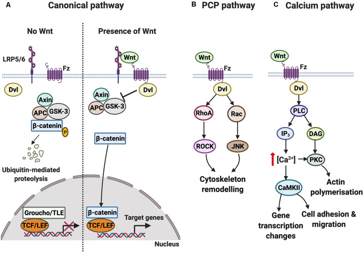
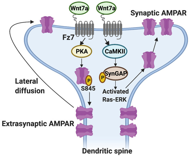
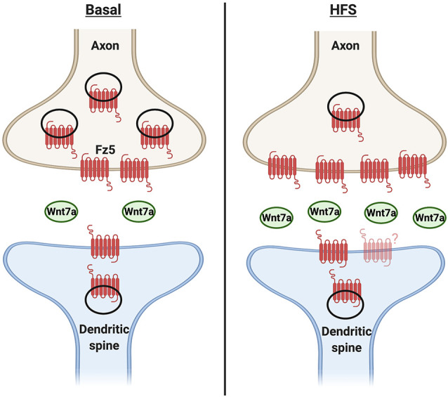
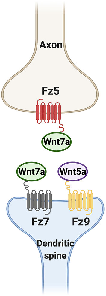
The formation of synapses is a tightly regulated process that requires the coordinated assembly of the presynaptic and postsynaptic sides. Defects in synaptogenesis during development or in the adult can lead to neurodevelopmental disorders, neurological disorders, and neurodegenerative diseases. In order to develop therapeutic approaches for these neurological conditions, we must first understand the molecular mechanisms that regulate synapse formation. The Wnt family of secreted glycoproteins are key regulators of synapse formation in different model systems from invertebrates to mammals. In this review, we will discuss the role of Wnt signaling in the formation of excitatory synapses in the mammalian brain by focusing on Wnt7a and Wnt5a, two Wnt ligands that play an in vivo role in this process. We will also discuss how changes in neuronal activity modulate the expression and/or release of Wnts, resulting in changes in the localization of surface levels of Frizzled, key Wnt receptors, at the synapse. Thus, changes in neuronal activity influence the magnitude of Wnt signaling, which in turn contributes to activity-mediated synapse formation.
Keywords: Wnt; development; frizzled; neuronal activity; synapse formation.
Copyright © 2021 Teo and Salinas.
Conflict of interest statement
The authors declare that the research was conducted in the absence of any commercial or financial relationships that could be construed as a potential conflict of interest.
Figures
Similar articles
-
Frizzled-5, a receptor for the synaptic organizer Wnt7a, regulates activity-mediated synaptogenesis.Development. 2010 Jul;137(13):2215-25. doi: 10.1242/dev.046722. Development. 2010. PMID: 20530549 Free PMC article.
-
Activity-mediated synapse formation a role for Wnt-Fz signaling.Curr Top Dev Biol. 2011;97:119-36. doi: 10.1016/B978-0-12-385975-4.00011-5. Curr Top Dev Biol. 2011. PMID: 22074604
-
Wnts acting through canonical and noncanonical signaling pathways exert opposite effects on hippocampal synapse formation.Neural Dev. 2008 Nov 5;3:32. doi: 10.1186/1749-8104-3-32. Neural Dev. 2008. PMID: 18986540 Free PMC article.
-
Wnts in action: from synapse formation to synaptic maintenance.Front Cell Neurosci. 2013 Nov 5;7:162. doi: 10.3389/fncel.2013.00162. Front Cell Neurosci. 2013. PMID: 24223536 Free PMC article. Review.
-
Postsynaptic assembly: a role for Wnt signaling.Dev Neurobiol. 2014 Aug;74(8):818-27. doi: 10.1002/dneu.22138. Epub 2013 Nov 15. Dev Neurobiol. 2014. PMID: 24105999 Free PMC article. Review.
Cited by
-
Gut neuroendocrine signaling regulates synaptic assembly in C. elegans.EMBO Rep. 2022 Aug 3;23(8):e53267. doi: 10.15252/embr.202153267. Epub 2022 Jun 24. EMBO Rep. 2022. PMID: 35748387 Free PMC article.
-
Patronin regulates presynaptic microtubule organization and neuromuscular junction development in Drosophila.iScience. 2024 Jan 18;27(2):108944. doi: 10.1016/j.isci.2024.108944. eCollection 2024 Feb 16. iScience. 2024. PMID: 38318379 Free PMC article.
-
Different strategies by distinct Wnt-signaling pathways in activating a nuclear transcriptional response.Curr Top Dev Biol. 2022;149:59-89. doi: 10.1016/bs.ctdb.2022.02.008. Epub 2022 Mar 4. Curr Top Dev Biol. 2022. PMID: 35606062 Free PMC article.
-
Anti-Müllerian Hormone Signal Transduction involved in Müllerian Duct Regression.Front Endocrinol (Lausanne). 2022 Jun 2;13:905324. doi: 10.3389/fendo.2022.905324. eCollection 2022. Front Endocrinol (Lausanne). 2022. PMID: 35721723 Free PMC article. Review.
-
Chinese acupuncture: A potential treatment for autism rat model via improving synaptic function.Heliyon. 2024 Aug 29;10(17):e37130. doi: 10.1016/j.heliyon.2024.e37130. eCollection 2024 Sep 15. Heliyon. 2024. PMID: 39286195 Free PMC article.
References
-
- Amaral M. D., Pozzo-Miller L. (2009). The dynamics of excitatory synapse formation on dendritic spines. Cellscience 5, 19–25. Available online at: https://pubmed.ncbi.nlm.nih.gov/20072712/ - PMC - PubMed
Grants and funding
LinkOut - more resources
Full Text Sources