Deletion of Clusterin Protects Cochlear Hair Cells against Hair Cell Aging and Ototoxicity
- PMID: 34194490
- PMCID: PMC8181089
- DOI: 10.1155/2021/9979157
Deletion of Clusterin Protects Cochlear Hair Cells against Hair Cell Aging and Ototoxicity
Abstract
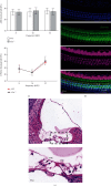
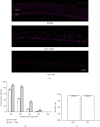
Hearing loss is a debilitating disease that affects 10% of adults worldwide. Most sensorineural hearing loss is caused by the loss of mechanosensitive hair cells in the cochlea, often due to aging, noise, and ototoxic drugs. The identification of genes that can be targeted to slow aging and reduce the vulnerability of hair cells to insults is critical for the prevention of sensorineural hearing loss. Our previous cell-specific transcriptome analysis of adult cochlear hair cells and supporting cells showed that Clu, encoding a secreted chaperone that is involved in several basic biological events, such as cell death, tumor progression, and neurodegenerative disorders, is expressed in hair cells and supporting cells. We generated Clu-null mice (C57BL/6) to investigate its role in the organ of Corti, the sensory epithelium responsible for hearing in the mammalian cochlea. We showed that the deletion of Clu did not affect the development of hair cells and supporting cells; hair cells and supporting cells appeared normal at 1 month of age. Auditory function tests showed that Clu-null mice had hearing thresholds comparable to those of wild-type littermates before 3 months of age. Interestingly, Clu-null mice displayed less hair cell and hearing loss compared to their wildtype littermates after 3 months. Furthermore, the deletion of Clu is protected against aminoglycoside-induced hair cell loss in both in vivo and in vitro models. Our findings suggested that the inhibition of Clu expression could represent a potential therapeutic strategy for the alleviation of age-related and ototoxic drug-induced hearing loss.
Copyright © 2021 Xiaochang Zhao et al.
Conflict of interest statement
The authors declare that the research was conducted in the absence of any commercial or financial relationships that could be construed as a potential conflict of interest.
Figures


Similar articles
-
Co-administration of cisplatin and furosemide causes rapid and massive loss of cochlear hair cells in mice.Neurotox Res. 2011 Nov;20(4):307-19. doi: 10.1007/s12640-011-9244-0. Epub 2011 Apr 1. Neurotox Res. 2011. PMID: 21455790 Free PMC article.
-
Scar formation in mice deafened with kanamycin and furosemide.Microsc Res Tech. 2016 Aug;79(8):766-72. doi: 10.1002/jemt.22695. Epub 2016 Jun 17. Microsc Res Tech. 2016. PMID: 27311812
-
Genetic and pharmacological intervention for treatment/prevention of hearing loss.J Commun Disord. 2008 Sep-Oct;41(5):421-43. doi: 10.1016/j.jcomdis.2008.03.004. Epub 2008 Mar 25. J Commun Disord. 2008. PMID: 18455177 Free PMC article. Review.
-
Systemic lipopolysaccharide induces cochlear inflammation and exacerbates the synergistic ototoxicity of kanamycin and furosemide.J Assoc Res Otolaryngol. 2014 Aug;15(4):555-70. doi: 10.1007/s10162-014-0458-8. Epub 2014 May 21. J Assoc Res Otolaryngol. 2014. PMID: 24845404 Free PMC article.
-
Genetic influences on susceptibility of the auditory system to aging and environmental factors.Scand Audiol Suppl. 1992;36:1-39. Scand Audiol Suppl. 1992. PMID: 1488615 Review.
Cited by
-
Emerging Mechanisms in the Pathogenesis of Menière's Disease: Evidence for the Involvement of Ion Homeostatic or Blood-Labyrinthine Barrier Dysfunction in Human Temporal Bones.Otol Neurotol. 2023 Dec 1;44(10):1057-1065. doi: 10.1097/MAO.0000000000004016. Epub 2023 Sep 20. Otol Neurotol. 2023. PMID: 37733989 Free PMC article.
-
Protein profile of mouse endolymph suggests a role in controlling cochlear homeostasis.iScience. 2024 Oct 19;27(11):111214. doi: 10.1016/j.isci.2024.111214. eCollection 2024 Nov 15. iScience. 2024. PMID: 39563888 Free PMC article.
-
Identification of common stria vascularis cellular alteration in sensorineural hearing loss based on ScRNA-seq.BMC Genomics. 2024 Feb 27;25(1):213. doi: 10.1186/s12864-024-10122-7. BMC Genomics. 2024. PMID: 38413848 Free PMC article.
-
Clusterin-mediated polarization of M2 macrophages: a mechanism of temozolomide resistance in glioblastoma stem cells.Stem Cell Res Ther. 2025 Mar 24;16(1):146. doi: 10.1186/s13287-025-04247-z. Stem Cell Res Ther. 2025. PMID: 40128761 Free PMC article.
-
The hair cell analysis toolbox is a precise and fully automated pipeline for whole cochlea hair cell quantification.PLoS Biol. 2023 Mar 22;21(3):e3002041. doi: 10.1371/journal.pbio.3002041. eCollection 2023 Mar. PLoS Biol. 2023. PMID: 36947567 Free PMC article.
References
-
- Vos T., Flaxman A. D., Naghavi M., et al. Years lived with disability (YLDs) for 1160 sequelae of 289 diseases and injuries 1990-2010: a systematic analysis for the Global Burden of Disease Study 2010. Lancet (London, England) 2012;380(9859):2163–2196. doi: 10.1016/S0140-6736(12)61729-2. - DOI - PMC - PubMed
Publication types
MeSH terms
Substances
LinkOut - more resources
Full Text Sources
Research Materials
Miscellaneous

