. 2021 May 29:2021:7615712.
doi: 10.1155/2021/7615712. eCollection 2021.

miRNA-6715-5p Inhibits Cellular Proliferation and Invasion in Colorectal Cancer by Directly Targeting CST4

Affiliations

miRNA-6715-5p Inhibits Cellular Proliferation and Invasion in Colorectal Cancer by Directly Targeting CST4

Ding Shi et al. J Oncol. .

Abstract

Background: Data on the correlation between CST4 and colorectal cancer (CRC) metastasis are scarce. The aim of this study was to analyze CST4 expression and investigate its biological roles and related microRNA- (miRNA-) mediated regulation in CRC.

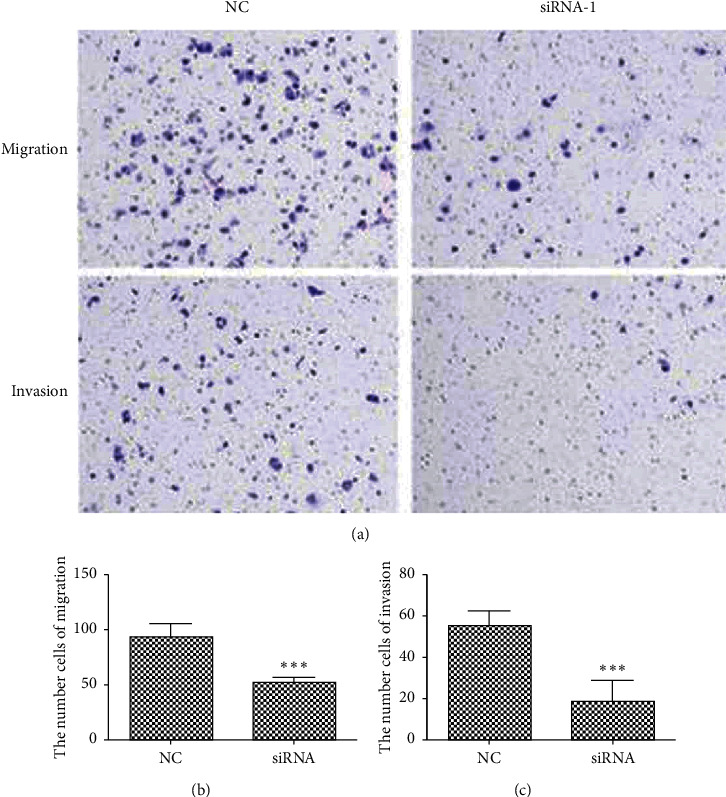
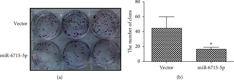
Methods: The expression of CST4 was examined in cancer tissues and their corresponding adjacent normal tissues from 40 gastric adenocarcinoma patients. The expression level of CST4 in specimens (cancer and normal tissues) was assessed through immunohistochemistry and/or quantitative polymerase chain reaction. miRNAs targeting CST4 in CRC were predicted by bioinformatics software. CST4 was knocked down in HCT116 cells and candidate miRNAs were transfected into HCT116 cells, and the effects of CST4 knockdown and miRNA transfection on cell proliferation and invasion were examined using CCK8, cell colony formation, and Transwell migration assays. Luciferase double-reporter assays were performed to verify the relationship between miRNA and CST4.

Results: The expression of CST4 in CRC tissues was significantly higher than that in normal paracancerous tissues, but the results for miRNA-6715-5p were opposite. Regardless of CST4 knockdown or miRNA-6715-5p overexpression, the proliferation and invasion ability of HCT116 cells decreased significantly. Luciferase double-reporter assays showed that the upregulation of miR-6715-5p significantly reduced the luciferase activities of the CST4 3'-UTR plasmid in HCT116 cells.

Conclusion: CST4 may be involved in CRC proliferation and metastasis. miRNA-6715-5p directly targets CST4 and negatively regulates its expression.


Conflict of interest statement

The authors declare that there are no conflicts of interest.

Figures

Figure 1
Figure 1
Detection of CST4 in colorectal cancer (CRC) cell lines by qPCR. The results showed that CST4 was expressed at the lowest level in HCT116 cells.
Figure 2
Figure 2
Expression of CST4 in colorectal cancer as examined by immunohistochemistry using an anti-CST4 antibody. CST4 positivity was brown or yellow in color by immunohistochemistry. Immunohistochemistry showed that CST4 staining in cancer tissue was stronger than that in paracancerous tissue. (a) Cancerous tissues; (b) paracancerous tissue.
Figure 3
Figure 3
Immunohistochemistry (IHC) and qPCR detection of CST4 expression in colorectal cancer (CRC) and adjacent tissues. (a) IHC; (b) qPCR. Compared with that in adjacent tissues, CST4 expression in CRC tissues was significantly upregulated (∗∗∗P < 0.001, ∗∗P < 0.01).
Figure 4
Figure 4
Detection of CST4 expression after a knockdown by qPCR and western blotting (WB). Compared with that in the negative control (NC), the expression of CST4 in human HCT116 cells transfected with siRNA (1, 2, and 3) was significantly decreased (∗∗∗P < 0.001, ∗∗P < 0.01). (a) qPCR; (b) WB. NC, irrelevant sequence; si1, siRNA1; si2, siRNA2; si3, siRNA3. The molecular weight of CST4 in cells was 16 kDa; the molecular weight of GAPDH cells was 42 kDa.
Figure 5
Figure 5
Cell viability after transfection of siCST4 at different time points based on CCK8 assays. Compared with that in the negative control (NC) at corresponding time points, CST4 siRNA-transfected cells showed a significant decrease in cell activity from the third day, and the degree of decrease increased with time (∗∗P < 0.01, ∗∗∗P < 0.001). NC, siRNA irrelevant sequence; siRNA, CST4 siRNA.
Figure 6
Figure 6
Colony formation assay results with CST4 knockdown and statistics. Compared with that in the negative control (NC), cell colony formation ability was significantly decreased after CST4 interference (∗∗P < 0.01). (a) Plate clone results; (b) statistics. NC, siRNA irrelevant sequence; siRNA, siCST4.
Figure 7
Figure 7
Migration and invasiveness of cells after knocking down CST4 as assessed by Transwell assays. Compared with that in the negative control (NC), the migration and invasion ability of cells decreased significantly after siRNA transfection (∗∗∗P < 0.001). (a) Migration and invasion results; (b) migration data; (c) invasion data. NC, siRNA irrelevant sequence; siRNA, siCST4.
Figure 8
Figure 8
Expression of CST4 after transfection with candidate miRNAs based on western blotting. Of nine candidate miRNAs, miRNA-6715-5p caused the most significant decrease in CST4 expression. The molecular weight of CST4 and GAPDH was 16 and 42 kDa, respectively. NC, miRNA irrelevant sequence.
Figure 9
Figure 9
Expression of CST4 after transfecting candidate miRNAs based on qPCR. Compared with that in the negative control (NC), of the nine candidate genes, both miRNA-4269 and miRNA-4472 caused the significant downregulation of CST4 expression (P < 0.05), whereas miRNA-6715-5p caused the most significant downregulation of CST4 expression (∗∗∗P < 0.001). NC, miRNA irrelevant sequence.
Figure 10
Figure 10
Luciferase double-reporter experiment to validate the expression of CST4 after transfecting candidate miRNAs. Compared with that in the negative control (NC), luminescence activity from the CST4 promoter in the groups treated with miRNA-4269 and miRNA-4472 decreased significantly (P < 0.05), and the most obvious decrease was with miRNA-6715-5p (∗∗∗P < 0.001). NC, miRNA irrelevant sequence.
Figure 11
Figure 11
Promoter activity analysis after cotransfection of CST4 and miRNA-6715-5p. Compared with that in the negative control (NC), the luminescence activity from the CST4 3′-UTR decreased significantly (∗∗∗P < 0.001) after cotransfection of CST4 and miRNA-6715-5p. NC, negative control; miR-6715, miR-6715-5p mimic; pmirGlo: pmirGlo no-load plasmid; CST4-3′-UTR, pmirGlo-CST4-3′-UTR.
Figure 12
Figure 12
Detection of miRNA-6715-5p in colorectal cancer (CRC) and adjacent tissues by qPCR. Compared with that in adjacent tissues, miRNA-6715-5p expression in CRC tissues was significantly downregulated (P < 0.05).
Figure 13
Figure 13
Cell viability at different time points after microRNA-6715-5p transfection based on CCK8 assays. Compared with that in the negative control (NC) at the corresponding time points, cell viability decreased significantly from the third day after miRNA-6715-5p transfection, and the degree of reduction increased with time (∗∗P < 0.01, ∗∗∗P < 0.001). NC, miRNA irrelevant sequence.
Figure 14
Figure 14
Colony formation after microRNA-6715-5p transfection. Compared with that of the cells with the empty plasmid, the colony formation ability of the cells transfected with miRNA-6715-5p was significantly reduced (P < 0.01). (a) Plate clone results. (b) statistics. Vector, empty plasmid.
Figure 15
Figure 15
Migration and invasiveness of cells after transfection with miRNA-6715-5p based on Transwell assays. Compared with that of the negative control (NC), the migration and invasion ability of cells decreased significantly after transfecting miRNA-6715-5p (n = 3, P < 0.05; ∗∗∗P < 0.001). (a) Migration and invasion results. (b) Migration data. (c) Invasion data. NC, empty plasmid.

Similar articles

Cited by

References

    1. Li T., Xiong Q., Zou Z., Lei X., Jiang Q., Liu D. Prognostic significance of cystatin SN associated nomograms in patients with colorectal cancer. Oncotarget. 2017;8(70):115153–115163. doi: 10.18632/oncotarget.23041. - DOI - PMC - PubMed
    1. Choi E. H., Kim J.-T., Kim J. H., et al. Upregulation of the cysteine protease inhibitor, cystatin SN, contributes to cell proliferation and cathepsin inhibition in gastric cancer. Clinica Chimica Acta. 2009;406(1-2):45–51. doi: 10.1016/j.cca.2009.05.008. - DOI - PubMed
    1. Yoneda K., Iida H., Endo H., et al. Identification of cystatin SN as a novel tumor marker for colorectal cancer. International Journal of Oncology. 2009;35:33–40. - PubMed
    1. Kim J.-T., Lee S.-J., Kang M. A., et al. Cystatin SN neutralizes the inhibitory effect of cystatin C on cathepsin B activity. Cell Death and Disease. 2013;4(12) doi: 10.1038/cddis.2013.485.e974 - DOI - PMC - PubMed
    1. Chen Y. F., Ma G., Cao X., et al. Overexpression of Cystatin SN positively affects survival of patients with surgically resected esophageal squamous cell carcinoma. BMC Surgury. 2013;13:p. 15. doi: 10.1186/1471-2482-13-15. - DOI - PMC - PubMed

LinkOut - more resources