

Intravascular leiomyomatosis with intracardiac extension: a toraco-abdominal approach
- PMID: 34194725
- PMCID: PMC8238396
- DOI: 10.1093/jscr/rjab249
Intravascular leiomyomatosis with intracardiac extension: a toraco-abdominal approach
Abstract
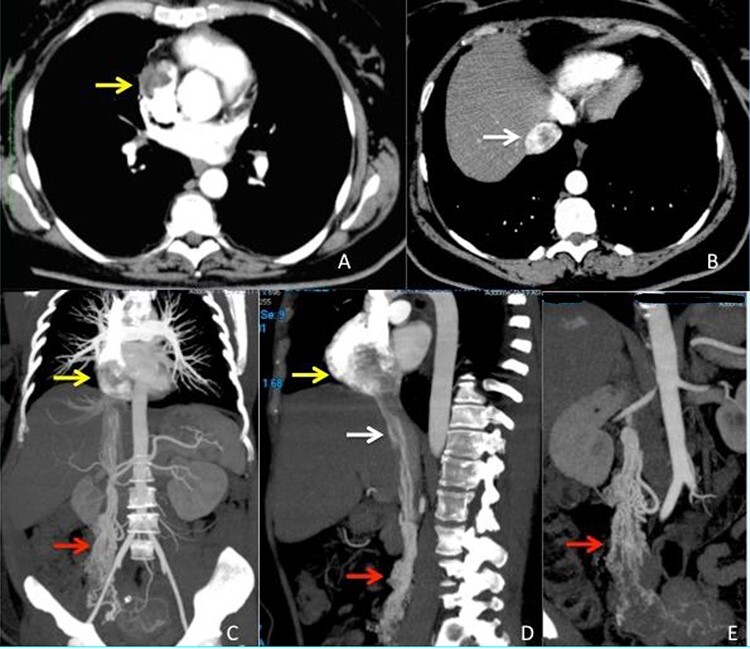
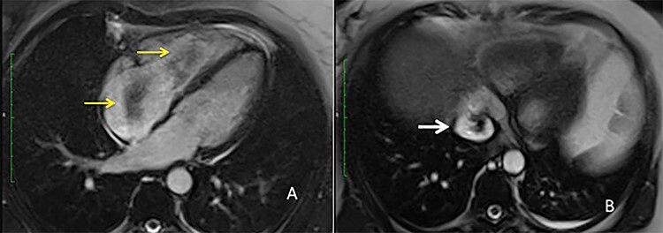
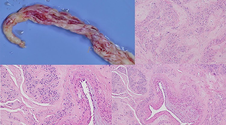
Intravenous leiomyomatosis is a rare nonmalignant tumor, which originates from the uterine smooth muscle cells and is usually confined to the pelvic venous system. Sometimes it can extend from the pelvis through the veins into the right side of the heart; this condition is named intracardiac leiomyomatosis (ICLM). To date few cases of these conditions have been described, the treatment is surgical, often challenging and usually multidisciplinary. In this paper are described the clinical presentation, the full radiologic study and surgical treatment of a case of ICLM that authors treated at their institution with thoraco-abdominal approach. Surgical removal of the ICLM is strongly recommended, because no recurrence has been reported, in our case at 7 years we did not observe recurrence of the disease.
Published by Oxford University Press and JSCR Publishing Ltd. All rights reserved. © The Author(s) 2021.
Figures
References
-
- Ma SQ, Bai CM, Yu XH, Huang OP, Lang JH, Li J. Clinical and pathological analyses of intravenous leiomyomatosis. Chin J Obstet Gynecol 2005;40:34–7. - PubMed
-
- Castelli P, Caronno R, Piggaretti G, Tozzi M. Intravenous uterine leiomyomatosis with right heart extension: successful two-stage surgical removal. Ann Vasc Surg 2006;20:405–7. - PubMed
-
- Mulvany NJ, Slavin JL, Ostor AG, Fortune D. Intravenous leiomyomatosis of the uterus: a clinicopathologic study of 22 cases. Int J Gynecol Pathol 1994;13:1–9. - PubMed
-
- Price JD, Anagnostopoulos C, Benvenisty A, Kothuru RK, Balaram SK. Intracardiac Extension of Intravenous Leiomyomatosis. Ann Thorac Surg 2017;103:e145–7. - PubMed
Publication types
LinkOut - more resources
Full Text Sources
