

Case Reports
doi: 10.1002/ccr3.4183.
eCollection 2021 Jun.
Laparoscopy-assisted immediate vaginal reconstruction with a vertical pedicled deep inferior epigastric perforator flap for primary melanoma of the vagina
Affiliations
- PMID: 34194773
- PMCID: PMC8223693
- DOI: 10.1002/ccr3.4183
Item in Clipboard
Case Reports
Laparoscopy-assisted immediate vaginal reconstruction with a vertical pedicled deep inferior epigastric perforator flap for primary melanoma of the vagina
Clin Case Rep.
.
Abstract
The vagina is a rare site for primary melanoma. Here, we report on a case of laparoscopy-assisted immediate vaginal reconstruction with vertical pedicled deep inferior epigastric perforator flap.
Keywords: DIEP; melanoma; treatment; vagina; vaginal reconstruction.
© 2021 Centre Hospitalier Universitaire de Brest. Clinical Case Reports published by John Wiley & Sons Ltd.
Conflict of interest statement
The authors report no conflicts of interest in relation to the present study.
Figures

Vaginal examination (A) of the PMV lesion. Black arrow: PMV lesion. MRI‐T2 sequence (B) of the PMV lesion. Black arrow: PMV lesion
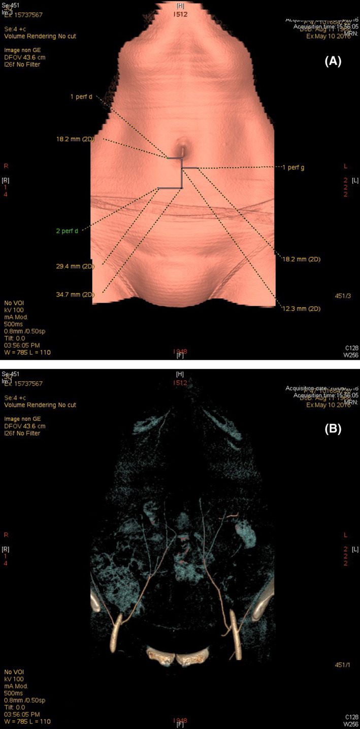
CT angiography of the deep inferior epigastric artery (A). A CT scan of the vascular network (B)

The inferior epigastric vessel transilluminated with the laparoscope's cold light source (A). EA: inferior epigastric artery

Laparoscopic view of the DIEP flap after its transfer to the pelvic cavity (A) EA: inferior epigastric artery UA: umbilical artery U: ureter. Laparoscopic view of the DIEP flap after its transfer to the pelvic cavity (A) EA: inferior epigastric artery UA: umbilical artery U: ureter. Laparoscopic view of the DIEP flap just before vaginal reconstruction (B) EA: inferior epigastric artery UA: umbilical artery U: ureter White arrow: The flap's two edges were sutured together, to give the flap a cylindrical shape

View of the neovagina after reconstruction by the DIEP flap. The length of the vagina is estimated by the length of the vaginal valve
Similar articles
-
The deep inferior epigastric artery perforator flap: a narrative review on its various uses in non-breast reconstruction.Ann Transl Med. 2023 Jan 31;11(2):130. doi: 10.21037/atm-22-2623. Epub 2022 Nov 29. Ann Transl Med. 2023. PMID: 36819501 Free PMC article. Review.
-
Vaginal reconstruction with pedicled vertical deep inferior epigastric perforator flap (diep) after pelvic exenteration. A consecutive case series.Gynecol Oncol. 2015 Sep;138(3):603-8. doi: 10.1016/j.ygyno.2015.06.031. Epub 2015 Jun 27. Gynecol Oncol. 2015. PMID: 26121919
-
Immediate vaginal reconstruction following pelvic exenteration using the pedicled vertical Deep Inferior Epigastric Perforator (DIEP) flap: A technical note.Ann Chir Plast Esthet. 2020 Jul;65(4):e1-e5. doi: 10.1016/j.anplas.2019.09.004. Epub 2020 Jun 11. Ann Chir Plast Esthet. 2020. PMID: 32536474
-
[Vaginal reconstruction with pedicled deep inferior epigastric perforator flap].Zhonghua Zheng Xing Wai Ke Za Zhi. 2009 Jan;25(1):8-10. Zhonghua Zheng Xing Wai Ke Za Zhi. 2009. PMID: 19408715 Chinese.
-
[Vaginal reconstruction after anterior pelvectomy by deep inferior epigastric perforator flap (DIEP)].Ann Urol (Paris). 2006 Jun;40(3):192-202. doi: 10.1016/j.anuro.2006.02.004. Ann Urol (Paris). 2006. PMID: 16869541 Review. French.
Cited by
-
The deep inferior epigastric artery perforator flap: a narrative review on its various uses in non-breast reconstruction.Ann Transl Med. 2023 Jan 31;11(2):130. doi: 10.21037/atm-22-2623. Epub 2022 Nov 29. Ann Transl Med. 2023. PMID: 36819501 Free PMC article. Review.
References
-
- Irvin WP, Bliss SA, Rice LW, Taylor PT, Andersen WA. Malignant melanoma of the vagina and locoregional control. Gynecol Oncol. 1998;71:476‐480. - PubMed
-
- Leitao MM, Cheng XI, Hamilton AL, et al. Gynecologic Cancer InterGroup (GCIG) consensus review for vulvovaginal melanomas. Int J Gynecol Cancer. 2014;24:S117‐S122. - PubMed
-
- Weinstock M. Malignant melanomas of the vulva and vagina in the United States: patterns of incidence and population‐based estimates of survival. Am J Obstet Gynecol. 1994;171:1225‐1230. - PubMed
-
- McLaughlin CC, Wu XC, Jemal A, Martin HJ, Roche LM, Chen VW. Incidence of noncutaneous melanomas in the U.S. Cancer. 2005;103:1000‐1007. - PubMed
-
- Ragnarsson‐Olding B, Johansson H, Rutqvist L‐E, Ringborg U. Malignant melanoma of the vulva and vagina: trends in incidence, age distribution, and long‐term survival among 245 consecutive cases in Sweden 1960–1984. Cancer. 1993;71:1893‐1897. - PubMed
Publication types
LinkOut - more resources
Full Text Sources
