. 2021 May 2;8(12):2100096.
doi: 10.1002/advs.202100096. eCollection 2021 Jun.

Topological Encoded Vector Beams for Monitoring Amyloid-Lipid Interactions in Microcavity

Affiliations

Topological Encoded Vector Beams for Monitoring Amyloid-Lipid Interactions in Microcavity

Chaoyang Gong et al. Adv Sci (Weinh). .

Abstract

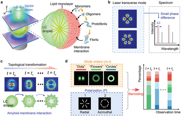
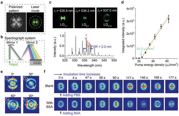
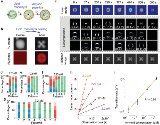
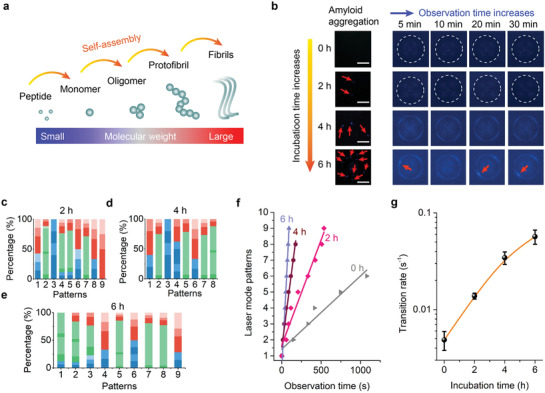
Lasers are the pillars of modern photonics and sensing. Recent advances in microlasers have demonstrated its extraordinary lasing characteristics suitable for biosensing. However, most lasers utilized lasing spectrum as a detection signal, which can hardly detect or characterize nanoscale structural changes in microcavity. Here the concept of amplified structured light-molecule interactions is introduced to monitor tiny bio-structural changes in a microcavity. Biomimetic liquid crystal droplets with self-assembled lipid monolayers are sandwiched in a Fabry-Pérot cavity, where subtle protein-lipid membrane interactions trigger the topological transformation of output vector beams. By exploiting Amyloid β (Aβ)-lipid membrane interactions as a proof-of-concept, it is demonstrated that vector laser beams can be viewed as a topology of complex laser modes and polarization states. The concept of topological-encoded laser barcodes is therefore developed to reveal dynamic changes of laser modes and Aβ-lipid interactions with different Aβ assembly structures. The findings demonstrate that the topology of vector beams represents significant features of intracavity nano-structural dynamics resulted from structured light-molecule interactions.

Keywords: amyloid‐lipid interaction; laser modes; liquid crystals; microcavity; topological structures; vector beams.


Conflict of interest statement

The authors declare no conflict of interest.

Figures

Figure 1
Figure 1
Concept of topological‐encoded laser barcode driven by molecular interactions. a) Schematic illustration of generating a vector beam driven by molecular interaction. Inset, illustration of mechanism. Aβ with different assemblies (monomers, oligomers, protofibrils, and fibrils) interact with the lipid monolayer coated on the LC droplet to trigger the topological transformation of the vector beam. b) Comparison of laser mode with conventional spectra interrogation. A small variation in intracavity phase can be detected by the topological transformation of transverse mode, however, the induced slight wavelength shift (Δλ) can hardly be recognized in spectrum. c) Schematic illustration of topological transformation in laser mode pattern. The molecular interaction on the LC droplet surface was amplified by the LC molecules and result in the topological transformation of laser mode. d) Illustration of the developed encoding rule. The observed laser mode pattern can be decomposed into Laguerre–Gaussian (LG) mode with different orders and polarization states, which was further encoded into the barcode.
Figure 2
Figure 2
Observation of transverse laser mode from LC droplet. a) Image of LC droplets taken under polarized optical microscopy (left panel) and the corresponding transverse laser mode pattern (right panel). The dashed lines indicate the radial director configuration, and the dashed circles indicate the size of the LC droplet. b) Schematic illustration of the hyperspectral imaging system. The laser mode pattern was dispersed by a diffractive grating according to its spectral components. c) The hyperspectral images (top) extracted from the laser mode in (a) and the corresponding laser emission spectrum (bottom). The spectrum is recorded at a pump energy density of 51.5 µJ mm−2. d) The spectrally integrated intensity with various pump energy densities. The error bars are obtained based on three times measurements. e) Polarization‐dependent laser mode patterns after passing through a linear polarizer with different angles. The direction of the maximum intensity (dashed line) agrees well with the orientation of the linear polarizer (arrow). f) Comparison of the temporal evolution of laser mode without (top) or with (bottom) 15 × 10–9 m BSA.
Figure 3
Figure 3
Topological transformation driven by Amyloid β‐lipid membrane interaction. a) Illustration of lipid monolayer coating and amyloid‐β (Aβ)‐lipid membrane interaction. b) Comparison of its corresponding polarized (PL) and fluorescence (FL) image before (left) and after (right) lipid monolayer coating. c) Comparison of the laser mode evolution and the polarized image. The laser mode decomposition results are also provided. The concentration of Aβ is 2.2 × 10⁻⁶ m. Topological laser mode barcode with Aβ concentration of d) 2.2 × 10–9 m, e) 22 × 10–9 m, f) 220 × 10–9 m, and g) 2.2 × 10⁻⁶ m, respectively. h) Comparison of laser mode temporal evolution under various Aβ concentrations. i) The transition rate as a function of Aβ concentrations.
Figure 4
Figure 4
Laser mode analysis of different amyloid‐β assembly structures interacting with lipid membrane. a) Illustration of Aβ self‐assembly structures. b) Left panel: Fluorescence microscopic images of Aβ‐lipid membrane interaction under different incubation times (0, 2, 4, 6 h) and observation times (5, 10, 20, 30 min). Scale bars, 10 µm. Topological laser barcodes converted from Aβ‐lipid membrane interaction under different incubation times of c) 2 h, d) 4 h, e) 6 h. f) Comparison of laser mode temporal evolution with different Aβ assembly structures interacting with lipid membranes. g) The transition rate as a function as incubation time.

Similar articles

Cited by

References

    1. Armani D. K., Kippenberg T. J., Spillane S. M., Vahala K. J., Nature 2003, 421, 925. - PubMed
    1. Zhang X., Cao Q. T., Wang Z., Liu Y. X., Qiu C. W., Yang L., Gong Q., Xiao Y. F., Nat. Photonics 2019, 13, 21.
    1. Armani A. M., Kulkarni R. P., Fraser S. E., Flagan R. C., Vahala K. J., Science 2007, 317, 783. - PubMed
    1. Baaske M. D., Foreman M. R., Vollmer F., Nat. Nanotechnol. 2014, 9, 933. - PubMed
    1. Chen W., Özdemir Ş. K., Zhao G., Wiersig J., Yang L., Nature 2017, 548, 192. - PubMed

Publication types

LinkOut - more resources