. 2021 Jun 5;24(6):102688.
doi: 10.1016/j.isci.2021.102688. eCollection 2021 Jun 25.

Human iPS cells engender corneal epithelial stem cells with holoclone-forming capabilities

Affiliations

Human iPS cells engender corneal epithelial stem cells with holoclone-forming capabilities

Shinya Watanabe et al. iScience. .

Abstract

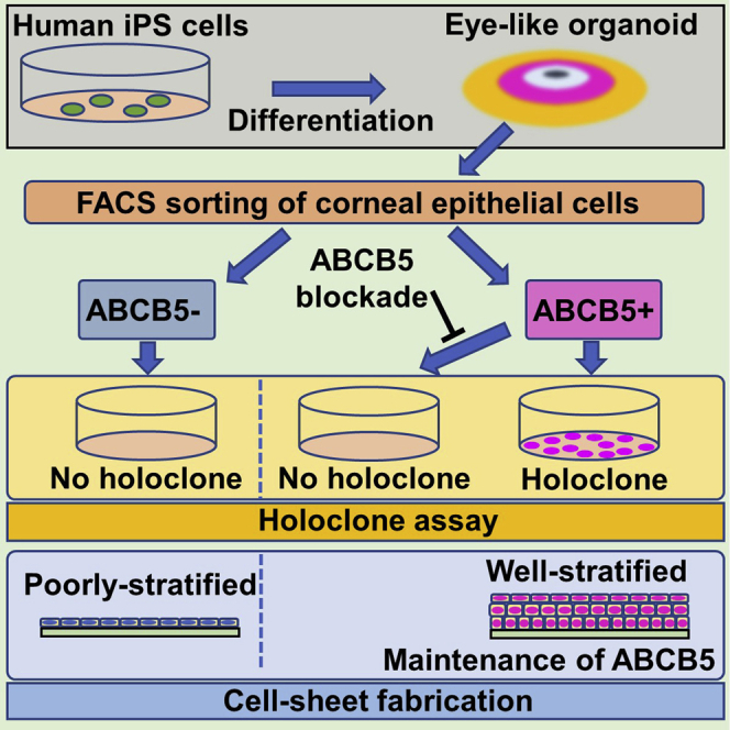
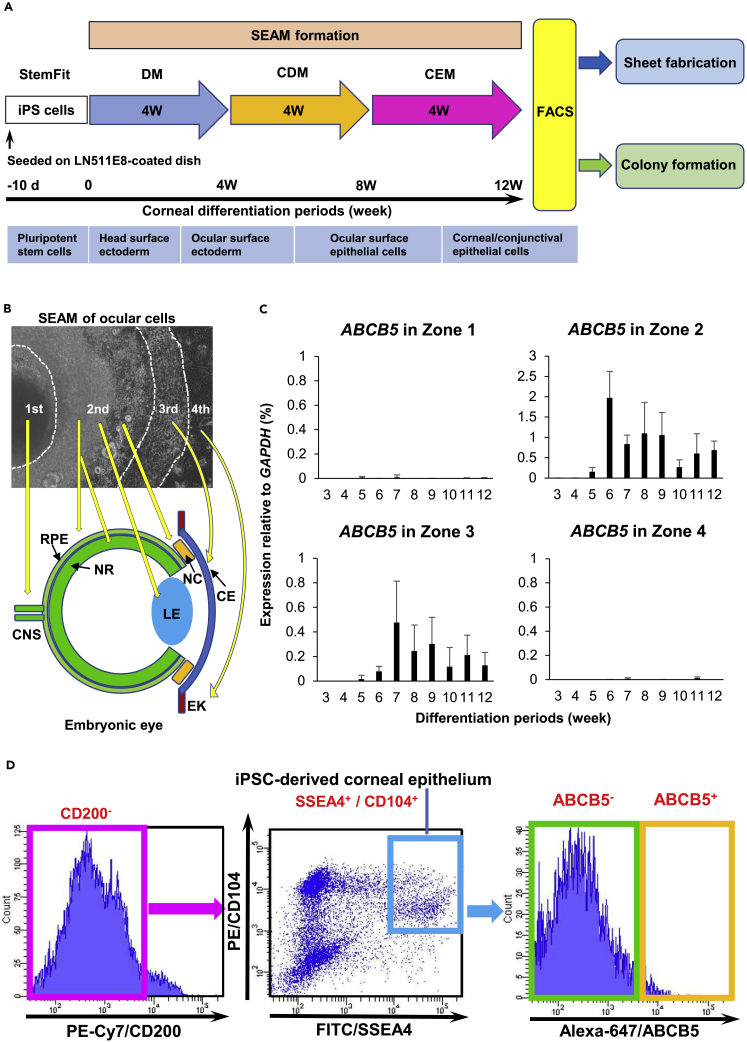
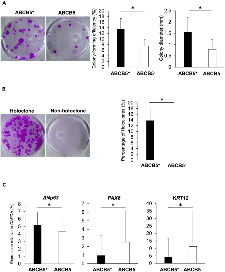
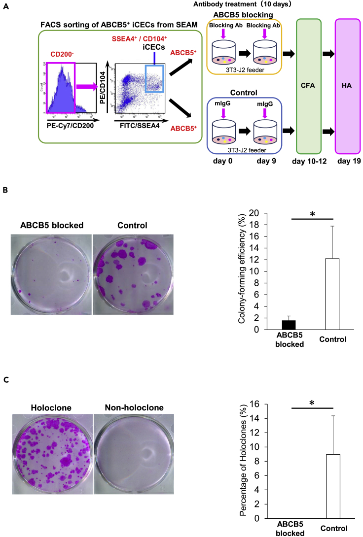
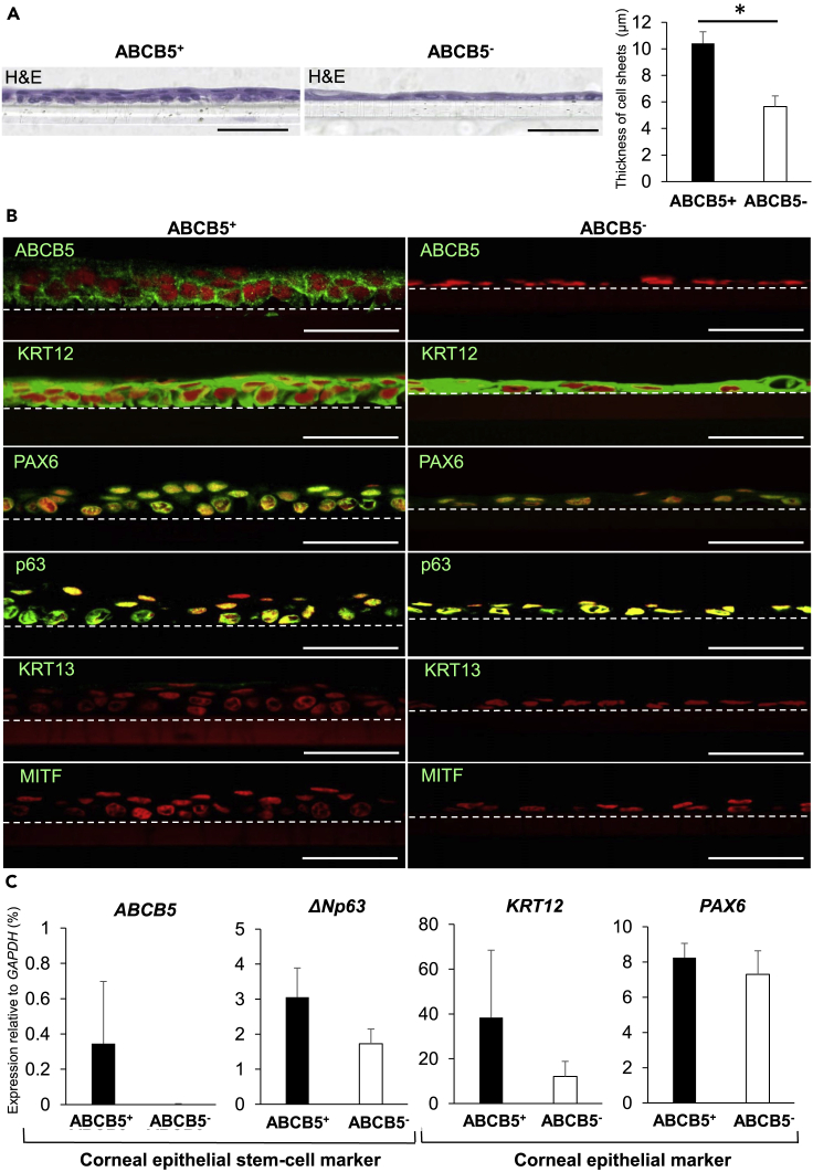
Human induced pluripotent stem cells (hiPSCs) can generate a multiplicity of organoids, yet no compelling evidence currently exists as to whether or not these contain tissue-specific, holoclone-forming stem cells. Here, we show that a subpopulation of cells in a hiPSC-derived corneal epithelial cell sheet is positive for ABCB5 (ATP-binding cassette, sub-family B, member 5), a functional marker of adult corneal epithelial stem cells. These cells possess remarkable holoclone-forming capabilities, which can be suppressed by an antibody-mediated ABCB5 blockade. The cell sheets are generated from ABCB5+ hiPSCs that first emerge in 2D eye-like organoids around six weeks of differentiation and display corneal epithelial immunostaining characteristics and gene expression patterns, including sustained expression of ABCB5. The findings highlight the translational potential of ABCB5-enriched, hiPSC-derived corneal epithelial cell sheets to recover vision in stem cell-deficient human eyes and represent the first report of holoclone-forming stem cells being directly identified in an hiPSC-derived organoid.

Keywords: Bioengineering; Cell biology; Stem cells research; Tissue engineering.


Conflict of interest statement

M.H.F., B.R.K., and N.Y.F. are inventors or co-inventors of US and international patents assigned to Brigham and Women's Hospital, Boston Children's Hospital, the Massachusetts Eye and Ear Infirmary, and/or the VA Boston Healthcare System, Boston, MA, licensed to TICEBA GmbH (Heidelberg, Germany) and RHEACELL GmbH & Co. KG (Heidelberg, Germany). M.H.F. serves as a scientific advisor for TICEBA GmbH and RHEACELL GmbH & Co. KG.

Figures

None
Graphical abstract
Figure 1
Figure 1
ABCB5+ corneal epithelial cells can be induced and isolated from hiPSCs (A) Strategy to generate and sort hiPSC-derived cells. CDM, corneal differentiation medium; CEM, corneal epithelium maintenance medium; DM, differentiation medium; d, day(s); w, week(s). (B) SEAM of hiPSCs mimics ocular development. CNS, central nervous system; NR, neuroretina; RPE, retinal pigment epithelium; NC, neural crest; LE; lens; CE, corneal epithelium; EK, epidermal keratinocyte. (C) ABCB5 expression in SEAM zones 1–4 during differentiation (each n = 5 independent experiments). Error bars are standard deviation. (D) Flow cytometric analysis of the differentiated SEAM. 0.71 ± 0.62% of hiPSC-derived corneal epithelial cells (SSEA4+/CD104+/CD200-) were ABCB5+. Data shown are representative of 58 independent cell sorting experiments.
Figure 2
Figure 2
hiPSC-derived ABCB5+ corneal epithelial cells have self-renewal potential (A) Representative colony-forming assays of seven independent experiments for ABCB5+/ABCB5- hiPSC-derived corneal epithelial cells plus colony-forming efficiency and colony diameter (left panel, n = 7 independent experiments; right panel, ABCB5+; n = 172 colonies: ABCB5-; n = 93 colonies from 4 independent experiments). (B) Representative images of a holoclone and a non-holoclone and percentage of holoclones (n = 4 independent experiments). See also Figure S1. (C), Gene expression analysis for stem cell marker ΔNp63 and differentiated corneal epithelium-related markers PAX6 and KRT12 in ABCB5+/ABCB5- colonies (ABCB5+; n = 131 colonies: ABCB5-; n = 55 colonies from 6 independent experiments). ∗p < 0.05, Wilcoxon signed-rank test. Error bars are standard deviation.
Figure 3
Figure 3
ABCB5 blockade inhibits self-renewal in hiPSC-derived ABCB5+ corneal epithelial cells (A) Schematic representation of mAb-mediated ABCB5 blocking followed by colony-forming assay (CFA) and holoclone assay (HA) for the ABCB5+ hiPSC-derived corneal epithelial cells (iCECs). Sorted ABCB5+ iCECs were treated with anti-ABCB5 blocking mAb or isotype control IgG for 10 consecutive days from the initial day of sorting. (B) Representative images of CFA after treatment with anti-ABCB5 blocking mAb or isotype control IgG (n = 4 independent experiments) and colony-forming efficiency after the treatments (n = 4 independent experiments). (C) Representative images of a holoclone and a non-holoclone after antibody treatment and percentage of holoclones after treatments with anti-ABCB5 blocking mAb or isotype control IgG (n = 4 independent experiments). ∗p < 0.05, Wilcoxon signed-rank test. Error bars are standard deviation.
Figure 4
Figure 4
Corneal epithelial cell sheets can be fabricated from hiPSC-derived ABCB5+/ABCB5- cells (A) Hematoxylin and eosin (H&E) staining of cell sheets fabricated from ABCB5+/ABCB5- hiPSC-derived corneal epithelial cells (iCECs). Scale bar, 50 μm. Comparative thickness of cell sheets fabricated from hiPSC-derived ABCB5+/ABCB5- cells. (n = 10 measurements from 1 experiment). ∗p < 0.05, Wilcoxon signed-rank test. Error bars are standard deviation. (B) Immunostaining for ABCB5 and ocular epithelial markers (green) in ABCB5+/ABCB5- iCEC-derived sheets. ABCB5, stem cell marker; KRT12, corneal epithelial marker, PAX6, ocular cell marker; p63, epithelial/surface ectodermal markers; KRT13, conjunctival epithelial marker; MITF, retinal pigment epithelium (RPE) marker. Nuclei, red. Scale bar, 50 μm. Dotted lines indicate border between cell sheets and culture inserts. (C) Gene expression analysis for corneal epithelial stem cell markers and corneal epithelium-related markers in ABCB5+/ABCB5- iCEC-derived sheets (n = 3 independent experiments). Error bars are standard deviation.

References

    1. Ang L.P., Sotozono C., Koizumi N., Suzuki T., Inatomi T., Kinoshita S. A comparison between cultivated and conventional limbal stem cell transplantation for Stevens-Johnson syndrome. Am. J. Ophthalmol. 2007;143:178–180. - PubMed
    1. Barrandon Y., Green H. Three clonal types of keratinocyte with different capacities for multiplication. Proc. Natl. Acad. Sci. U S A. 1987;84:2302–2306. - PMC - PubMed
    1. Chen K.G., Szakács G., Annereau J., Rouzaud F., Liang X., Valencia J.C., Nagineni C.N., Hooks J.J., Hearing V.J., Gottesman M.M. Principal expression of two mRNA isoforms (ABCB 5alpha and ABCB 5beta) of the ATP-binding cassette transporter gene ABCB 5 in melanoma cells and melanocytes. Pigment Cell Res. 2005;18:102–112. - PMC - PubMed
    1. Frank N.Y., Pendse S.S., Lapchak P.H., Margaryan A., Shlain D., Doeing C., Sayegh M.H., Frank M.H. Regulation of progenitor cell fusion by ABCB5 P-glycoprotein, a novel human ATP-binding cassette transporter. J. Biol. Chem. 2003;278:47156–47165. - PubMed
    1. Gain P., Jullienne R., He Z., Aldossary M., Acquart S., Cognasse F., Thuret G. Global survey of corneal transplantation and eye banking. JAMA Ophthalmol. 2016;134:167–173. - PubMed

LinkOut - more resources