. 2021 May 24;2(2):100544.
doi: 10.1016/j.xpro.2021.100544. eCollection 2021 Jun 18.

Preparation of the intact rodent organ of Corti for RNAscope and immunolabeling, confocal microscopy, and quantitative analysis

Affiliations

Preparation of the intact rodent organ of Corti for RNAscope and immunolabeling, confocal microscopy, and quantitative analysis

Daniel O J Reijntjes et al. STAR Protoc. .

Abstract

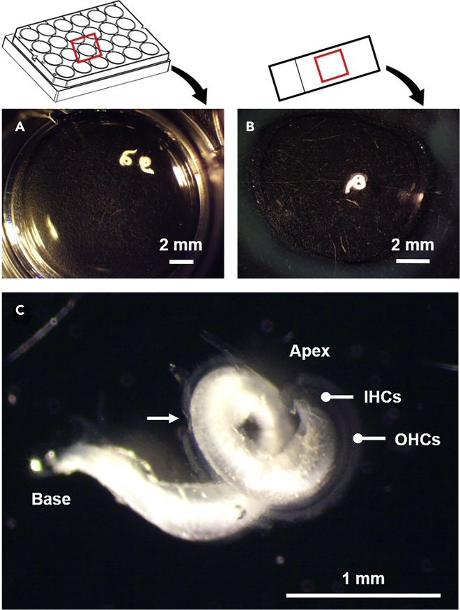
This protocol describes the preparation of the mouse organ of Corti for RNAscope, immunolabeling, confocal microscopy, and quantitative image analysis to examine transcript and protein localization, sensory hair cells, and synapses. This protocol can be applied to mice and other rodents (juvenile and adult) and can be adapted for other techniques, including electrophysiology and RNA sequencing. This protocol features minimal tissue processing to preserve viability for downstream assays, while isolating the organ of Corti is the most challenging step. For additional details on the use and execution of this protocol, please refer to McLean et al. (2009); Schuth et al. (2014); Lingle et al. (2019); Pyott et al. (2020).

Keywords: Antibody; In Situ Hybridization; Microscopy; Molecular/Chemical Probes; Neuroscience.


Conflict of interest statement

The authors declare no competing interests.

Figures

None
Graphical abstract
Figure 1
Figure 1
Isolation and preparation of the inner ear for fixation (A) The lines of cut (dashed lines) and region of the temporal bone (red box) containing the inner ear are indicated. (B) The inner ear, comprising the cochlea and vestibule, is attached to the temporal bone (TB) adjacent to the tentorium cerebelli (TC). The auditory and vestibular nerves exit the cochlea and vestibule, respectively, and fuse to form the vestibulocochlear nerve (CNVIII). (C) The cochlea of the isolated inner (now rotated 180°around the vertical axis in the orientation shown in A) shows several anatomical features, including the round window (RW) and oval window (OW). The apex of the cochlea (containing the portion of the organ of Corti, or auditory sensory epithelium, responsive to the lowest frequencies of sound) is marked. The mouse cochlea contains an apical, middle, and basal turn, distinguished by the two dashed lines. (D) A small hole in the bone overlaying the apex has been made to allow better access to fixative and is outlined with a dashed oval. The dissection is shown from a mouse aged 6 weeks old.
Figure 2
Figure 2
Fixed and isolated organs of Corti for RNAscope and immunostaining Isolated organs of Corti are processed in 24-well plates filed with solution (A) or on slides in solution contained within a circle-shaped hydrophobic barrier (B). (C) The organ of Corti spirals from the apex to base. Even at low magnification, the orderly arrangement of the inner hair cells (IHCs) and outer hair cells (OHCs) in rows spiraling the length of the organ of Corti is visible. The arrow marks a section of the organ of Corti that has been damaged during dissection.
Figure 3
Figure 3
Mounting organs of Corti for microscopy (A) Organs of Corti are placed into individual drops of mounting medium. (B) To prevent turns of the organ of Corti from overlapping, the organ of Corti is cut into two or three pieces before mounting with a glass coverslip (as shown here). The apex and base are indicated. The arrow marks an air bubble caught inadvertently under the coverslip.
Figure 4
Figure 4
Epifluorescent imaging of the immunostained and mounted organ of Corti (A) Three lower magnification epifluorescent micrographs of the apical, middle, and basal turns of the immunostained and mounted organ of Corti have been combined into a single digital image to overlay a tonotopic map (yellow). (B) A higher magnification epifluorescent micrograph reveals the spiral ganglion neurons (SGNs, circled) and rows of inner hair cells (IHCs) and outer hair cells (OHCs). In both A and B, the organ of Corti is immunostained with rabbit anti-calretinin and goat anti-Rabbit (488).
Figure 5
Figure 5
Z-projection through a stack of confocal micrographs of an organ of Corti With identification of the appropriate markers, a variety of structures can be identified in precisely defined tonotopic regions of the immunostained organ of Corti. (A) Organ of Corti isolated from Thy1-YFP-16 transgenic mice (Feng et al., 2000) express yellow fluorescent protein (green) in the myelinated neurons contacting the inner hair cells (IHCs) and outer hair cells (OHCs). IHCs and OHCs are immunostained with rabbit anti-MYO6 (red). A video of a 3D rotation is available as a supplemental video (Methods video S1). (B) Organ of Corti showing abundant expression of Lrrc52 transcript (green) detected by RNAscope in the inner hair cells (IHCs). IHCs are immunostained with rabbit anti-calretinin (red).
Figure 6
Figure 6
Quantitative 2D image analysis using ImageJ (A) Z-projection through a stack of confocal micrographs of an organ of Corti immunostained with anti-CTBP2 to label IHC nuclei (larger, fainter green immunolabels with example marked by an asterisk) and pre-synaptic ribbons (smaller, brighter green labels immunolabels with example marked by an arrow) bright. (B) Grayscale image of A for subsequent processing. (C) Black and white (inverted) image of B for subsequent processing. (D) Detected puncta with properties (e.g., diameters and surface areas) available for export. Complete instructions are available from the lead contact.
Figure 7
Figure 7
Quantitative 3D image analysis using Imaris and custom script (A) 3D reconstruction of a stack of confocal micrographs of an organ of Corti immunostained with mouse anti-CTBP2 to label IHC nuclei and pre-synaptic ribbons (green) and rabbit anti- GluR2/3 to label postsynaptic glutamate receptor clusters (red). (B) Spots detection of IHC nuclei (yellow spheres), pre-synaptic ribbons (green squares) and post-synaptic glutamate receptor clusters (red squares) for counting and spatial (x, y, z) localization. (C) Surface detection of pre-synaptic ribbons (green surfaces) and post-synaptic glutamate receptor clusters (red square surfaces) for volume analysis and spatial (x, y, z) localization.
Figure 8
Figure 8
Non-specific immunoreactivity (problem 3) Z-projections through a stack of confocal micrographs of an organ of Corti immunostained with anti-CTBP2 (green) to label presynaptic ribbons and anti-GluR2 (red) to label postsynaptic terminals. In panels (A and B), GluA2 immunoreactivity is clustered (A) and adjacent CTBP2-labeled presynaptic presynaptic ribbons (B). In contrast, in panels (C and D), GluA2 immunoreactivity is diffuse and not clearly clustered to the postsynaptic terminals (C) adjacent the CTBP2-labeled presynaptic ribbons (D).
Figure 9
Figure 9
Regions of the organ of Corti are damaged or missing or not mounted flatly (Problem 5) A higher magnification epifluorescent micrograph reveals the three rows of outer hair cells (OHCs) immunolabeled with rabbit anti-prestin. The dashed boxes show regions where OHCs are missing. The solid box marks a region where OHCs are not mounted flatly.

References

    1. Barone C.M., Douma S., Reijntjes D.O.J., Browe B.M., Koppl C., Klump G., Park T.J., Pyott S.J. Altered cochlear innervation in developing and mature naked and Damaraland mole rats. J. Comp. Neurol. 2019;527:2302–2316. - PMC - PubMed
    1. Braude J.P., Vijayakumar S., Baumgarner K., Laurine R., Jones T.A., Jones S.M., Pyott S.J. Deletion of Shank1 has minimal effects on the molecular composition and function of glutamatergic afferent postsynapses in the mouse inner ear. Hear. Res. 2015;321:52–64. - PMC - PubMed
    1. Feng G., Mellor R.H., Bernstein M., Keller-Peck C., Nguyen Q.T., Wallace M., Nerbonne J.M., Lichtman J.W., Sanes J.R. Imaging neuronal subsets in transgenic mice expressing multiple spectral variants of GFP. Neuron. 2000;28:41–51. - PubMed
    1. Goutman J.D., Pyott S.J. Whole-Cell Patch-Clamp Recording of Mouse and Rat Inner Hair Cells in the Intact Organ of Corti. Methods Mol. Biol. 2016;1427:471–485. - PubMed
    1. Greguske E.A., Llorens J., Pyott S.J. Assessment of cochlear toxicity in response to chronic 3,3’-iminodipropionitrile in mice reveals early and reversible functional loss that precedes overt histopathology. Arch. Toxicol. 2021;95:1003–1021. - PMC - PubMed

Publication types

LinkOut - more resources