

Optimized immunofluorescence staining protocol for imaging germinal centers in secondary lymphoid tissues of vaccinated mice
- PMID: 34195671
- PMCID: PMC8233161
- DOI: 10.1016/j.xpro.2021.100499
Optimized immunofluorescence staining protocol for imaging germinal centers in secondary lymphoid tissues of vaccinated mice
Abstract
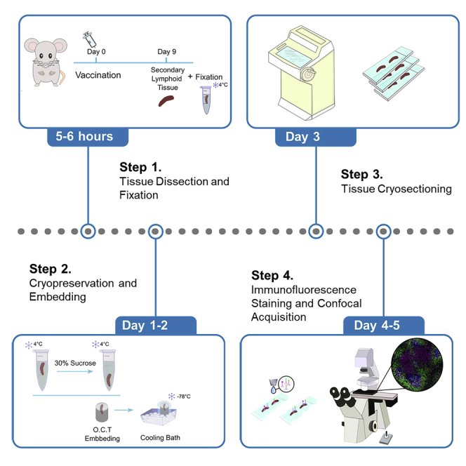
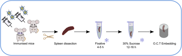
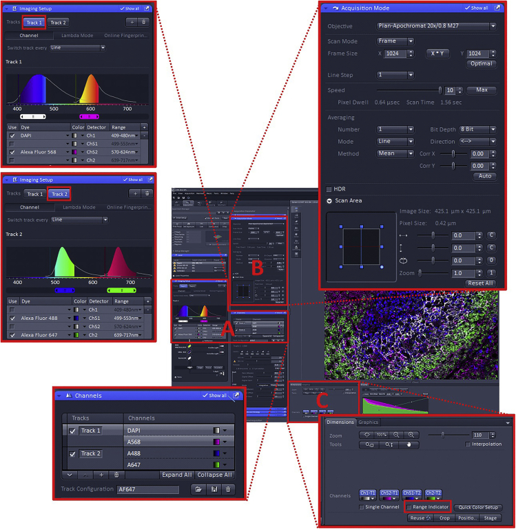
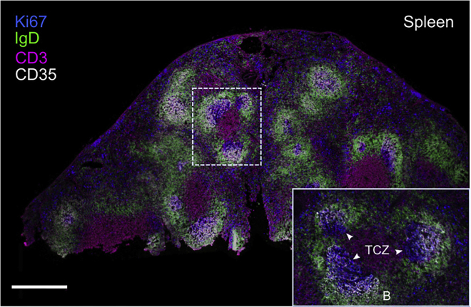
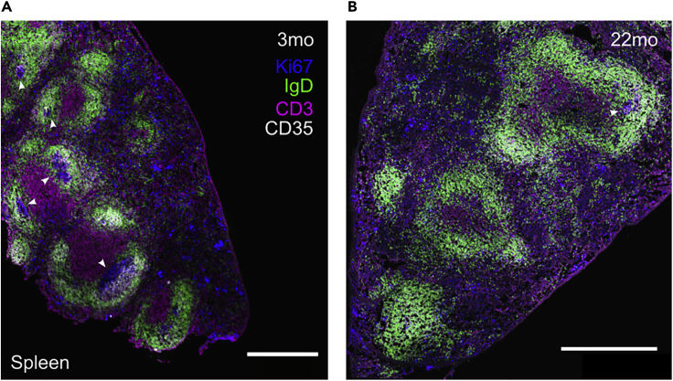
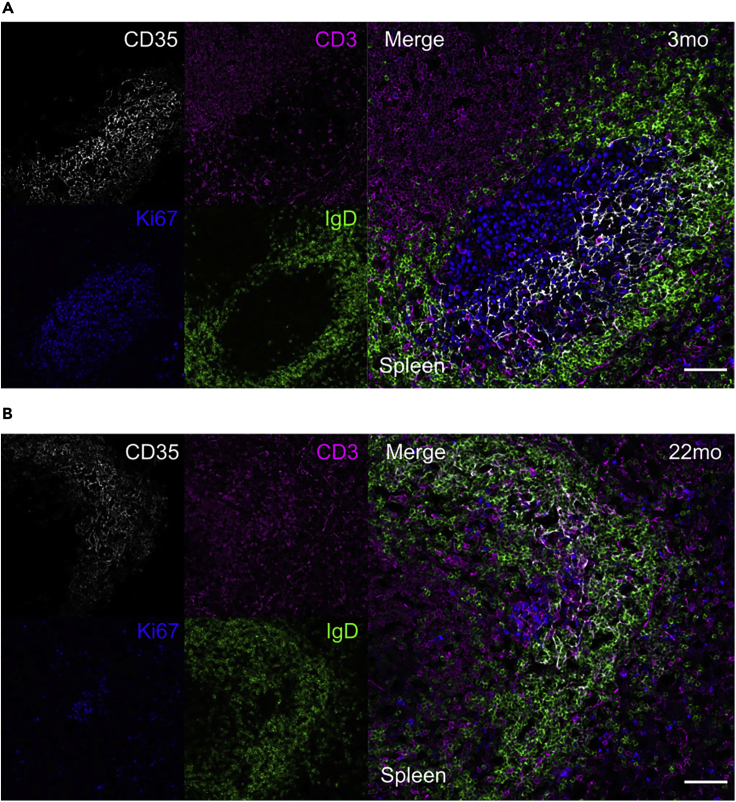
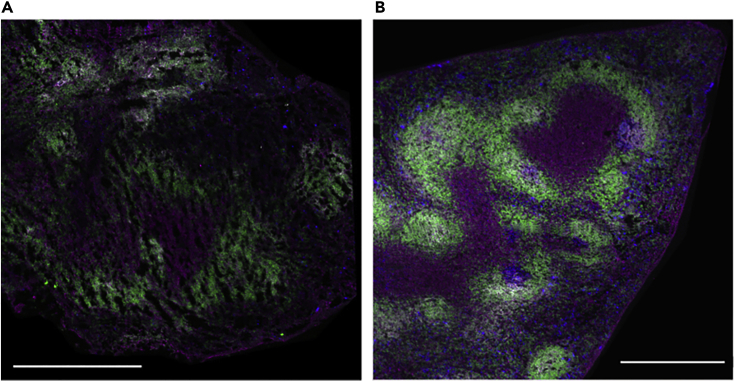
Location of immune cells that form the germinal center reaction within secondary lymphoid tissues can be characterized using confocal microscopy. Here, we present an optimized immunofluorescence staining protocol to image germinal center structures in fixed/frozen spleen sections from ChAdOx1 nCoV-19 immunized mice. This protocol can be adapted to identify other cell types within secondary lymphoid tissues. For complete information on the generation and use of this protocol to examine immune responses to the COVID vaccine ChAdOx1 nCoV-19, please refer to Silva-Cayetano et al. (2020).
Keywords: Antibody; Cell Biology; Immunology; Microbiology; Microscopy; Model Organisms.
© 2021 The Authors.
Conflict of interest statement
The authors declare no competing interests.
Figures
References
-
- Jonkman J., Brown C.M., Wright G.D., Anderson K.I., North A.J. Tutorial: guidance for quantitative confocal microscopy. Nat. Protoc. 2020;15:1585–1611. - PubMed
-
- Mclean I.W., Nakane P.K. Periodate-lysine-paraformaldehyde fixative a new fixative for immunoelectron microscopy. J. Histochem. Cytochem. 1974;22:1077–1083. - PubMed
-
- Silva-Cayetano A., Foster W.S., Innocentin S., Belij-Rammerstorfer S., Spencer A.J., Burton O.T., Fra-Bido S., Le Lee J., Thakur N., Conceicao C. A booster dose enhances immunogenicity of the COVID-19 vaccine candidate ChAdOx1 nCoV-19 in aged mice. Med (N Y) 2020 doi: 10.1016/j.medj.2020.12.006. - DOI - PMC - PubMed
Publication types
MeSH terms
Substances
LinkOut - more resources
Full Text Sources
Medical
