. 2021 Aug;59(2):59.
doi: 10.3892/ijo.2021.5239. Epub 2021 Jul 1.

Cancer‑associated fibroblast‑induced M2‑polarized macrophages promote hepatocellular carcinoma progression via the plasminogen activator inhibitor‑1 pathway

Affiliations

Cancer‑associated fibroblast‑induced M2‑polarized macrophages promote hepatocellular carcinoma progression via the plasminogen activator inhibitor‑1 pathway

Shuhai Chen et al. Int J Oncol. 2021 Aug.

Abstract

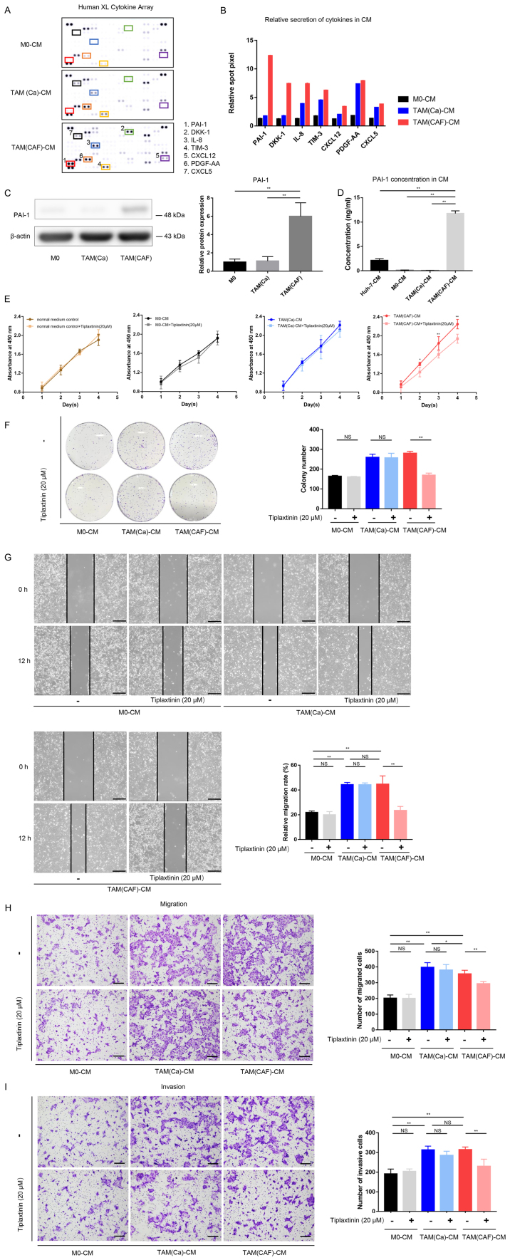
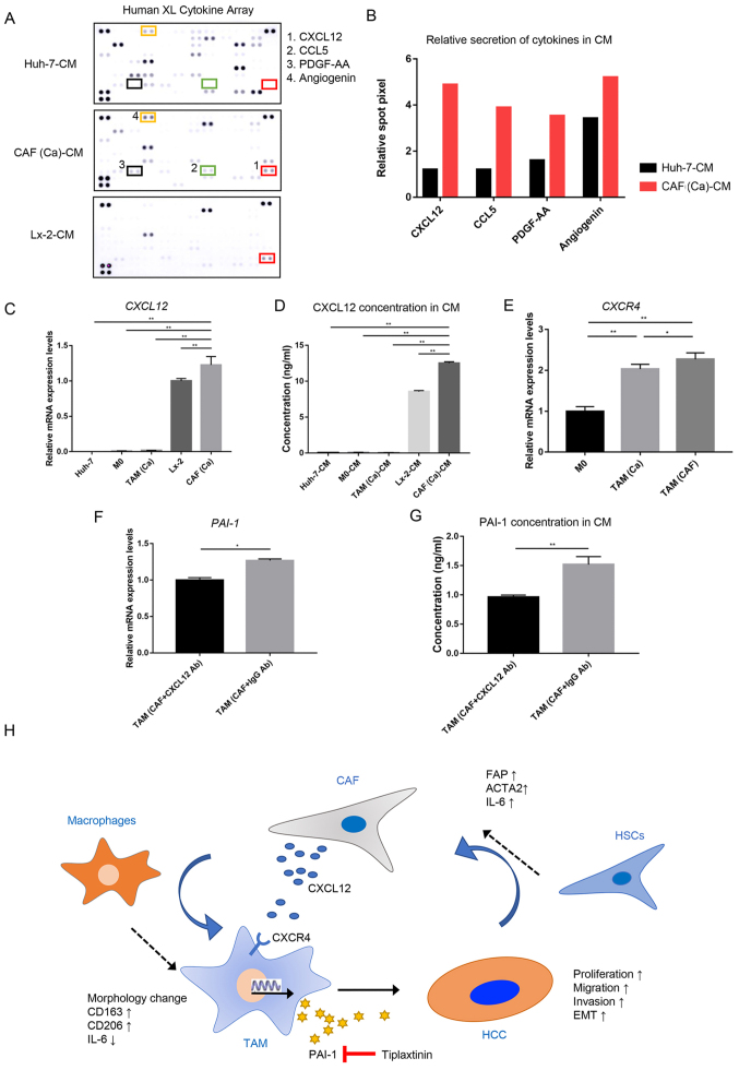
Targeting the tumor stroma is an important strategy in cancer treatment. Cancer‑associated fibroblasts (CAFs) and tumor‑associated macrophages (TAMs) are two main components in the tumor microenvironment (TME) in hepatocellular carcinoma (HCC), which can promote tumor progression. Plasminogen activator inhibitor‑1 (PAI‑1) upregulation in HCC is predictive of unfavorable tumor behavior and prognosis. However, the crosstalk between cancer cells, TAMs and CAFs, and the functions of PAI‑1 in HCC remain to be fully investigated. In the present study, macrophage polarization and key paracrine factors were assessed during their interactions with CAFs and cancer cells. Cell proliferation, wound healing and Transwell and Matrigel assays were used to investigate the malignant behavior of HCC cells in vitro. It was found that cancer cells and CAFs induced the M2 polarization of TAMs by upregulating the mRNA expression levels of CD163 and CD206, and downregulating IL‑6 mRNA expression and secretion in the macrophages. Both TAMs derived from cancer cells and CAFs promoted HCC cell proliferation and invasion. Furthermore, PAI‑1 expression was upregulated in TAMs after being stimulated with CAF‑conditioned medium and promoted the malignant behavior of the HCC cells by mediating epithelial‑mesenchymal transition. CAFs were the main producer of C‑X‑C motif chemokine ligand 12 (CXCL12) in the TME and CXCL12 contributed to the induction of PAI‑1 secretion in TAMs. In conclusion, the results of the present study suggested that CAFs promoted the M2 polarization of macrophages and induced PAI‑1 secretion via CXCL12. Furthermore, it was found that PAI‑1 produced by the TAMs enhanced the malignant behavior of the HCC cells. Therefore, these factors may be targets for inhibiting the crosstalk between tumor cells, CAFs and TAMs.

Keywords: cancer‑associated fibroblasts; epithelial‑mesenchymal transition; hepatocellular carcinoma; plasminogen activator inhibitor‑1; tumor‑associated macrophages.


Conflict of interest statement

The authors declare that they have no competing interests.

Figures

Figure 1
Figure 1
Cancer cells and CAFs induce M2 polarization in human macrophages. (A) Schematic representation of the generation of M0, CAFs and the three types of TAMs. (B) Schematic representation of anti-CXCL12 treatment in CAF-induced TAMs. (C) After treatment with CM from the Huh-7 cells, the gene expression levels of the CAFs markers were detected in the Lx-2 cells using RT-qPCR analysis. (D) Typical morphological changes in the M0 macrophages and different types of TAMs are shown following staining with H&E. Scale bar, 100 µm. (E) Gene expression levels of M2-polarized macrophage markers were detected in M0 macrophages and different types of TAMs using RT-qPCR analysis. (F) Secretion of IL-6 in the CM of M0 and different types of TAMs. *P<0.05;**P<0.01. CAF, cancer-associated fibroblast; CM, conditioned medium; RT-qPCR, reverse transcription-quantitative PCR; TAM, tumor-associated macrophage; PMA, phorbol-12-myristate-13-acetate; M0, macrophages; FAP, fibroblast activation protein; ACTA2, actin α-2; CXCL12, C-X-C motif chemokine ligand 12; Ab, antibody.
Figure 2
Figure 2
TAM(Ca) and TAM(CAF) promote HCC cell malignant behavior in vitro. (A) Cell Counting Kit-8 and (B) colony formation assay results demonstrated the effect of different types of TAMs on the proliferation of the Huh-7 cells. *P<0.05, **P<0.01, #P<0.05 and ##P<0.01 vs. M0-CM. (C) Wound healing (scale bar, 400 µm) and (D) Transwell and (E) Matrigel assays (scale bar, 200 µm) indicated that TAM(Ca) and TAM(CAF) promoted Huh-7 cell migration and invasion compared with that in the M0. **P<0.01. CAF, cancer-associated fibroblast; TAM, tumor-associated macrophage; CM, conditioned medium; M0, macrophages; NS, not significant.
Figure 3
Figure 3
PAI-1 is the key factor secreted by TAMs following CAF-CM stimulation and promotes tumor malignant behavior in vitro. (A) Original image and (B) spot pixel value from a cytokine array revealed the profiles of paracrine factors in the M0-CM, TAM (Ca)-CM and TAM(CAF)-CM. (C) Western blot and (D) ELISA results indicated that PAI-1 was upregulated in the TAM(CAF) group compared with that in the M0 and TAM(Ca) groups. (E) Cell Counting Kit-8 and (F) colony formation assays showed that the inhibition of PAI-1 in TAM(CAF)-CM suppressed the enhanced proliferation of Huh-7 cells. (G) Wound healing (scale bar, 400 µm) and (H) Transwell and (I) Matrigel (scale bar, 200 µm) assays indicated that the inhibition of PAI-1 in the TAM(CAF)-CM group suppressed the enhanced migration and invasion of the Huh-7 cells. *P<0.05; **P<0.01. CAF, cancer-associated fibroblast; CM, conditioned medium; CXCL12, C-X-C motif chemokine ligand 12; CXCL5, C-X-C motif chemokine ligand 5; PAI-1, plasminogen activator inhibitor-1; PDGF-AA, platelet derived growth factor-AA; TAM, tumor-associated macrophage; TIM-3, T-cell immunoglobulin mucin 3; M0, macrophages; NS, not significant.
Figure 4
Figure 4
PAI-1 promotes HCC malignant behavior by mediating EMT. (A) Cell Counting Kit-8, (B) wound healing (scale bar, 400 µm), (C) Transwell and (D) Matrigel (scale bar, 200 µm) assays demonstrated the direct effects of PAI-1 on tumor cell malignant behavior. (E) EMT-related gene expression was detected using reverse transcription-quantitative PCR in the Huh-7 cells. (F) Western blot analysis revealed the protein expression levels of E-cadherin, N-cadherin, Snail and Twist1 in the Huh-7 cells under the specified treatment conditions. *P<0.05;**P<0.01. CAF, cancer-associated fibroblast; CM, conditioned medium; PAI-1, plasminogen activator inhibitor-1; Snail, snail family transcriptional repressor 1; TAM, tumor-associated macrophage; Twist1, twist family bHLH transcription factor 1; NS, not significant.
Figure 5
Figure 5
CAF-derived CXCL12 induces the secretion of PAI-1 in TAM(CAF). (A) Original image and (B) spot pixel value from a cytokine array to identify the different patterns of molecules in cancer-CM and CAFs-CM. CXCL12 gene expression and its secretion were increased in CAFs compared with that in the Huh-7 and Lx-2 cells following (C) RT-qPCR and (D) ELISA. (E) CXCR4 gene expression in M0, TAM(Ca) and TAM(CAF) was analyzed using RT-qPCR. (F) RT-qPCR and (G) ELISA results demonstrated that the gene expression level and secretion of PAI-1 were decreased in TAM(CAF) after CXCL12 neutralization in CAF-CM, respectively. (H) Schematic representation of the proposed interactions between HCC, TAMs and CAFs. *P<0.05; **P<0.01. CAF, cancer-associated fibroblast; CM, conditioned medium; CCL5, C-C motif chemokine ligand 5; CXCL12, C-X-C motif chemokine ligand 12; CXCR4, C-X-C Motif chemokine receptor 4; HCC, hepatocellular carcinoma; HSCs, hepatic stellate cells; PAI-1, plasminogen activator inhibitor-1; PDGF-AA, platelet derived growth factor-AA; RT-qPCR, reverse transcription-quantitative PCR; TAM, tumor-associated macrophage; Ab, antibody.

References

    1. Metcalf KJ, Alazzeh A, Werb Z, Weaver VM. Leveraging microenvironmental synthetic lethalities to treat cancer. J Clin Invest. 2021;131:e143765. doi: 10.1172/JCI143765. - DOI - PMC - PubMed
    1. Quail DF, Joyce JA. Microenvironmental regulation of tumor progression and metastasis. Nat Med. 2013;19:1423–1437. doi: 10.1038/nm.3394. - DOI - PMC - PubMed
    1. Bejarano L, Jordāo MJ, Joyce JA. Therapeutic targeting of the tumor microenvironment. Cancer Discov. 2021;11:933–959. doi: 10.1158/2159-8290.CD-20-1808. - DOI - PubMed
    1. Comito G, Giannoni E, Segura CP, Barcellos-de-Souza P, Raspollini MR, Baroni G, Lanciotti M, Serni S, Chiarugi P. Cancer-associated fibroblasts and M2-polarized macrophages synergize during prostate carcinoma progression. Oncogene. 2014;33:2423–2431. doi: 10.1038/onc.2013.191. - DOI - PubMed
    1. Andersson P, Yang Y, Hosaka K, Zhang Y, Fischer C, Braun H, Liu S, Yu G, Liu S, Beyaert R, et al. Molecular mechanisms of IL-33-mediated stromal interactions in cancer metastasis. JCI Insight. 2018;3:e122375. doi: 10.1172/jci.insight.122375. - DOI - PMC - PubMed

MeSH terms

Substances