

Endothelial-Mesenchymal Transition in Cardiovascular Disease
- PMID: 34196216
- PMCID: PMC8387428
- DOI: 10.1161/ATVBAHA.121.313788
Endothelial-Mesenchymal Transition in Cardiovascular Disease
Abstract
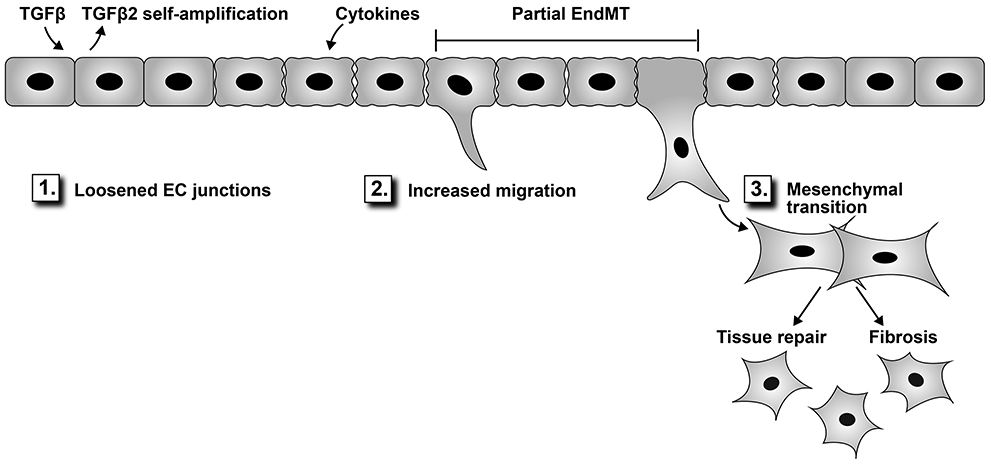
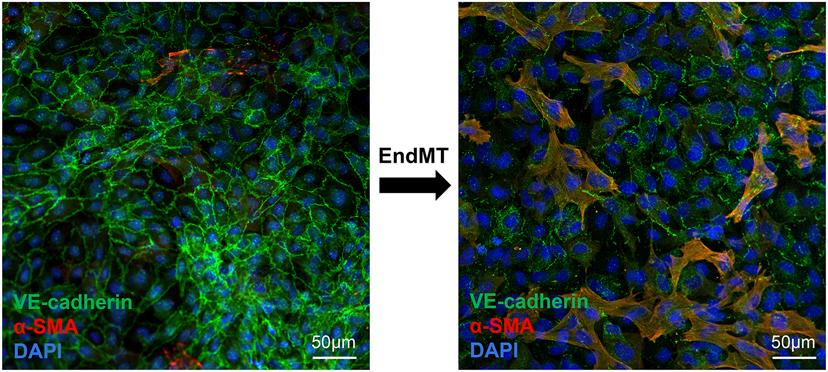
Endothelial-to-mesenchymal transition is a dynamic process in which endothelial cells suppress constituent endothelial properties and take on mesenchymal cell behaviors. To begin the process, endothelial cells loosen their cell-cell junctions, degrade the basement membrane, and migrate out into the perivascular surroundings. These initial endothelial behaviors reflect a transient modulation of cellular phenotype, that is, a phenotypic modulation, that is sometimes referred to as partial endothelial-to-mesenchymal transition. Loosening of endothelial junctions and migration are also seen in inflammatory and angiogenic settings such that endothelial cells initiating endothelial-to-mesenchymal transition have overlapping behaviors and gene expression with endothelial cells responding to inflammatory signals or sprouting to form new blood vessels. Reduced endothelial junctions increase permeability, which facilitates leukocyte trafficking, whereas endothelial migration precedes angiogenic sprouting and neovascularization; both endothelial barriers and quiescence are restored as inflammatory and angiogenic stimuli subside. Complete endothelial-to-mesenchymal transition proceeds beyond phenotypic modulation such that mesenchymal characteristics become prominent and endothelial functions diminish. In proadaptive, regenerative settings the new mesenchymal cells produce extracellular matrix and contribute to tissue integrity whereas in maladaptive, pathologic settings the new mesenchymal cells become fibrotic, overproducing matrix to cause tissue stiffness, which eventually impacts function. Here we will review what is known about how TGF (transforming growth factor) β influences this continuum from junctional loosening to cellular migration and its relevance to cardiovascular diseases.
Keywords: cardiovascular disease; endothelial cells; mitral valve insufficiency; phenotype; transforming growth factor.
Figures
References
-
- Markwald RR, Fitzharris TP and Manasek FJ. Structural development of endocardial cushions. Am J Anat. 1977;148:85–119. - PubMed
-
- Markwald RR, Fitzharris TP and Smith WN. Structural analysis of endocardial cytodifferentiation. Developmental Biology. 1975;42:160–80. - PubMed
-
- Ursoli Ferreira F, Eduardo Botelho Souza L, Hassibe Thome C, Tomazini Pinto M, Origassa C, Salustiano S, Marcel Faca V, Olsen Camara N, Kashima S and Tadeu Covas D. Endothelial Cells Tissue-Specific Origins Affects Their Responsiveness to TGF-beta2 during Endothelial-to-Mesenchymal Transition. Int J Mol Sci. 2019;20. - PMC - PubMed
-
- Nakano A, Nakano H, Smith KA and Palpant NJ. The developmental origins and lineage contributions of endocardial endothelium. Biochim Biophys Acta. 2016;1863:1937–47. - PubMed
Publication types
MeSH terms
Grants and funding
LinkOut - more resources
Full Text Sources
