

Immunohistochemical evaluation of immune cell infiltration in canine gliomas
- PMID: 34196247
- PMCID: PMC11404454
- DOI: 10.1177/03009858211023946
Immunohistochemical evaluation of immune cell infiltration in canine gliomas
Abstract
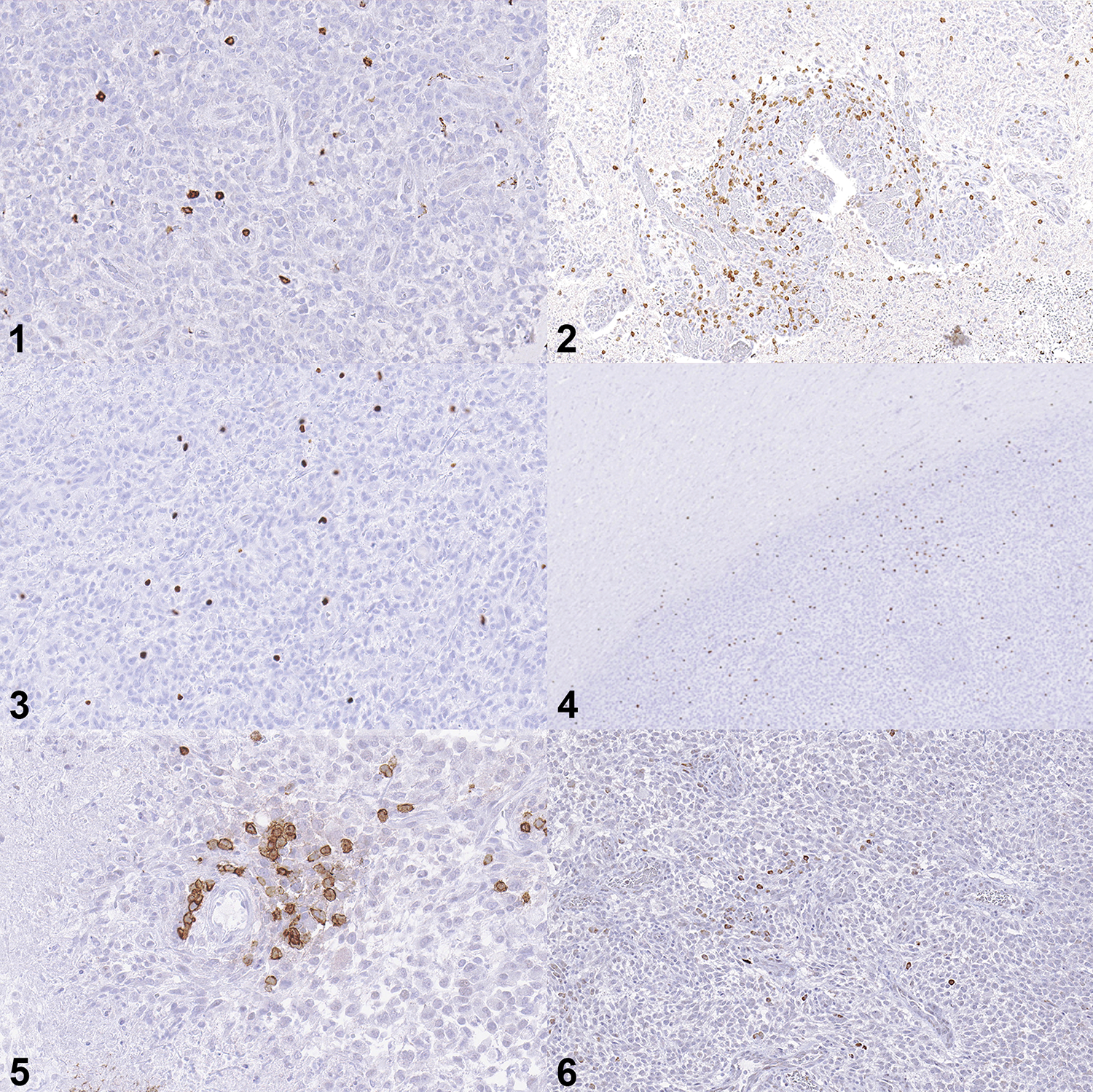
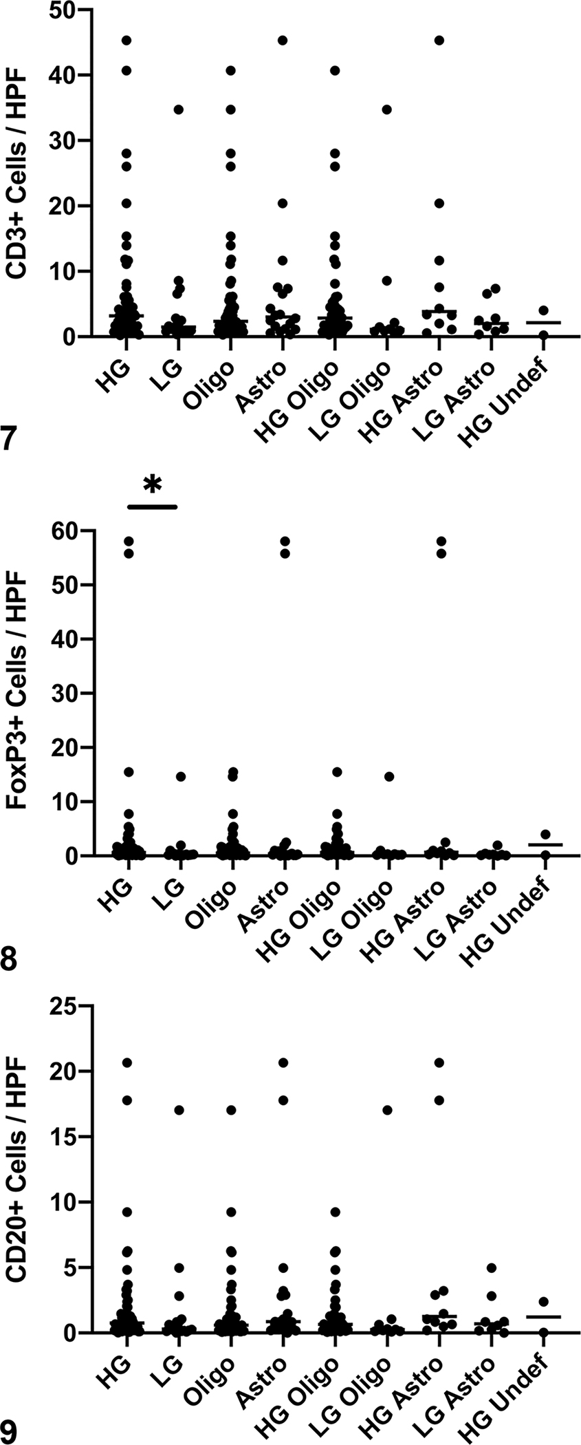
Evasion of the immune response is an integral part of the pathogenesis of glioma. In humans, important mechanisms of immune evasion include recruitment of regulatory T cells (Tregs) and polarization of macrophages toward an M2 phenotype. Canine glioma has a robust immune cell infiltrate that has not been extensively characterized. The purpose of this study was to determine the distribution of immune cells infiltrating spontaneous intracranial canine gliomas. Seventy-three formalin-fixed, paraffin-embedded tumor samples were evaluated using immunohistochemistry for CD3, forkhead box 3 (FOXP3), CD20, Iba1, calprotectin (Mac387), CD163, and indoleamine 2,3-dioxygenase (IDO). Immune cell infiltration was present in all tumors. Low-grade and high-grade gliomas significantly differed in the numbers of FoxP3+ cells, Mac387+ cells, and CD163+ cells (P = .006, .01, and .01, respectively). Considering all tumors, there was a significant increase in tumor area fraction of CD163 compared to Mac387 (P < .0001), and this ratio was greater in high-grade tumors than in low-grade tumors (P = .005). These data warrant further exploration into the roles of macrophage repolarization or Treg interference therapy in canine glioma.
Keywords: astrocytoma; brain; cancer; dogs; immunohistochemistry; immunology; lymphocytes; macrophages; oligodendroglioma.
Conflict of interest statement
DECLARATION OF CONFLICTING INTERESTS
The authors declared no potential conflicts of interest with respect to the research, authorship, or publication of this article.
Figures


References
Publication types
MeSH terms
Substances
Grants and funding
LinkOut - more resources
Full Text Sources
Research Materials
