Exploiting DNA Endonucleases to Advance Mechanisms of DNA Repair
- PMID: 34198612
- PMCID: PMC8232306
- DOI: 10.3390/biology10060530
Exploiting DNA Endonucleases to Advance Mechanisms of DNA Repair
Abstract
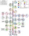
The earliest methods of genome editing, such as zinc-finger nucleases (ZFN) and transcription activator-like effector nucleases (TALENs), utilize customizable DNA-binding motifs to target the genome at specific loci. While these approaches provided sequence-specific gene-editing capacity, the laborious process of designing and synthesizing recombinant nucleases to recognize a specific target sequence, combined with limited target choices and poor editing efficiency, ultimately minimized the broad utility of these systems. The discovery of clustered regularly interspaced short palindromic repeat sequences (CRISPR) in Escherichia coli dates to 1987, yet it was another 20 years before CRISPR and the CRISPR-associated (Cas) proteins were identified as part of the microbial adaptive immune system, by targeting phage DNA, to fight bacteriophage reinfection. By 2013, CRISPR/Cas9 systems had been engineered to allow gene editing in mammalian cells. The ease of design, low cytotoxicity, and increased efficiency have made CRISPR/Cas9 and its related systems the designer nucleases of choice for many. In this review, we discuss the various CRISPR systems and their broad utility in genome manipulation. We will explore how CRISPR-controlled modifications have advanced our understanding of the mechanisms of genome stability, using the modulation of DNA repair genes as examples.
Keywords: CRISPR; base excision repair; gene editing; homologous recombination; microhomology-mediated end-joining; mismatch repair; non-homologous end-joining.
Conflict of interest statement
RWS is a scientific consultant for Canal House Biosciences, LLC. The authors state that there is no conflict of interest.
Figures


Similar articles
-
Prescription of Controlled Substances: Benefits and Risks.2025 Jul 6. In: StatPearls [Internet]. Treasure Island (FL): StatPearls Publishing; 2025 Jan–. 2025 Jul 6. In: StatPearls [Internet]. Treasure Island (FL): StatPearls Publishing; 2025 Jan–. PMID: 30726003 Free Books & Documents.
-
Advancing crop disease resistance through genome editing: a promising approach for enhancing agricultural production.Front Genome Ed. 2024 Jun 26;6:1399051. doi: 10.3389/fgeed.2024.1399051. eCollection 2024. Front Genome Ed. 2024. PMID: 38988891 Free PMC article. Review.
-
Evolution of Prime Editing Systems: Move Forward to the Treatment of Hereditary Diseases.Curr Gene Ther. 2025;25(1):46-61. doi: 10.2174/0115665232295117240405070809. Curr Gene Ther. 2025. PMID: 38623982 Review.
-
Identification of Mycobacterium tuberculosis intracellular survival-related virulence factors via CRISPR-based eukaryotic-like secretory protein mutant library screen.Microbiol Spectr. 2025 Aug 5;13(8):e0076725. doi: 10.1128/spectrum.00767-25. Epub 2025 Jun 12. Microbiol Spectr. 2025. PMID: 40503836 Free PMC article.
-
Target-dependent nickase activities of the CRISPR-Cas nucleases Cpf1 and Cas9.Nat Microbiol. 2019 May;4(5):888-897. doi: 10.1038/s41564-019-0382-0. Epub 2019 Mar 4. Nat Microbiol. 2019. PMID: 30833733 Free PMC article.
Cited by
-
CRISPR/Cas9-Induced Knockout of miR-24 Reduces Cholesterol and Monounsaturated Fatty Acid Content in Primary Goat Mammary Epithelial Cells.Foods. 2022 Jul 7;11(14):2012. doi: 10.3390/foods11142012. Foods. 2022. PMID: 35885255 Free PMC article.
-
dCas9 binding inhibits the initiation of base excision repair in vitro.DNA Repair (Amst). 2022 Jan;109:103257. doi: 10.1016/j.dnarep.2021.103257. Epub 2021 Nov 20. DNA Repair (Amst). 2022. PMID: 34847381 Free PMC article.
-
Tips, Tricks, and Potential Pitfalls of CRISPR Genome Editing in Saccharomyces cerevisiae.Front Bioeng Biotechnol. 2022 May 30;10:924914. doi: 10.3389/fbioe.2022.924914. eCollection 2022. Front Bioeng Biotechnol. 2022. PMID: 35706506 Free PMC article. Review.
References
Publication types
Grants and funding
- R01 CA238061/CA/NCI NIH HHS/United States
- P01 ES028949/ES/NIEHS NIH HHS/United States
- ES028949/NH/NIH HHS/United States
- R01 ES014811/ES/NIEHS NIH HHS/United States
- R01ES030084/ES/NIEHS NIH HHS/United States
- ES014811/NH/NIH HHS/United States
- R01 ES030084/ES/NIEHS NIH HHS/United States
- CA238061/NH/NIH HHS/United States
- AG069740/NH/NIH HHS/United States
- GRANT11998991/U.S. Department of Defense
- R01 CA148629/CA/NCI NIH HHS/United States
- R44 ES032522/ES/NIEHS NIH HHS/United States
- ES032522/NH/NIH HHS/United States
- U01 ES029518/ES/NIEHS NIH HHS/United States
- ES029518/NH/NIH HHS/United States
- R35ES031708/ES/NIEHS NIH HHS/United States
- NSF-1841811/National Science Foundation
- R01 AG069740/AG/NIA NIH HHS/United States
- CA148629/NH/NIH HHS/United States
LinkOut - more resources
Full Text Sources

