. 2021 Jun 5;22(11):6100.
doi: 10.3390/ijms22116100.

The Propensity of the Human Liver to Form Large Lipid Droplets Is Associated with PNPLA3 Polymorphism, Reduced INSIG1 and NPC1L1 Expression and Increased Fibrogenetic Capacity

Affiliations

The Propensity of the Human Liver to Form Large Lipid Droplets Is Associated with PNPLA3 Polymorphism, Reduced INSIG1 and NPC1L1 Expression and Increased Fibrogenetic Capacity

Flaminia Ferri et al. Int J Mol Sci. .

Abstract

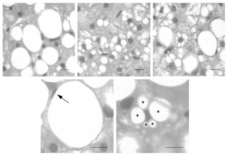
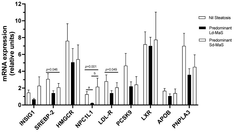
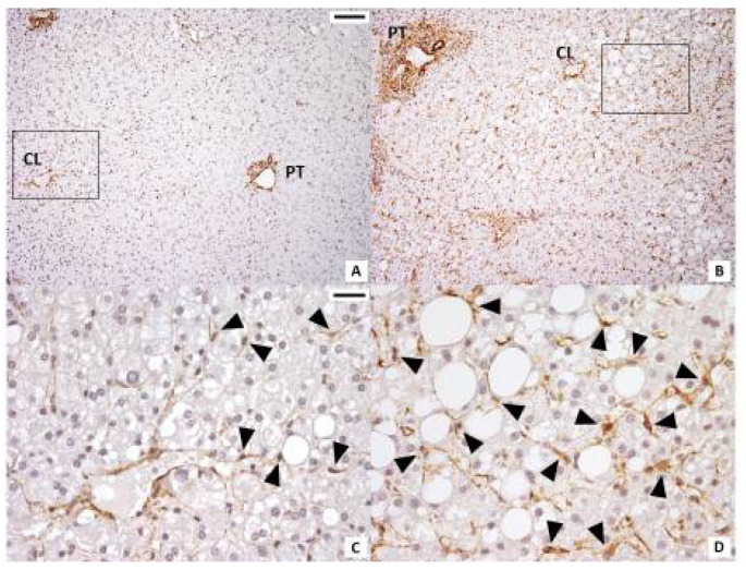
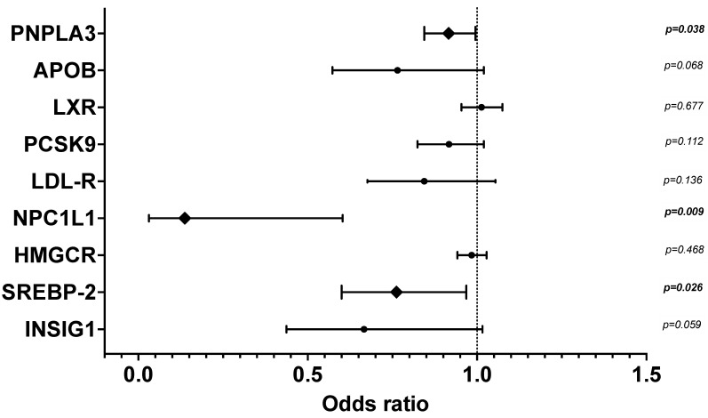
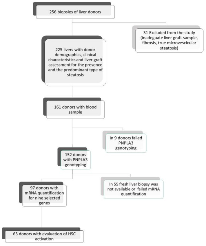
In nonalcoholic steatohepatitis animal models, an increased lipid droplet size in hepatocytes is associated with fibrogenesis. Hepatocytes with large droplet (Ld-MaS) or small droplet (Sd-MaS) macrovesicular steatosis may coexist in the human liver, but the factors associated with the predominance of one type over the other, including hepatic fibrogenic capacity, are unknown. In pre-ischemic liver biopsies from 225 consecutive liver transplant donors, we retrospectively counted hepatocytes with Ld-MaS and Sd-MaS and defined the predominant type of steatosis as involving ≥50% of steatotic hepatocytes. We analyzed a donor Patatin-like phospholipase domain-containing protein 3 (PNPLA3) rs738409 polymorphism, hepatic expression of proteins involved in lipid metabolism by RT-PCR, hepatic stellate cell (HSC) activation by α-SMA immunohistochemistry and, one year after transplantation, histological progression of fibrosis due to Hepatitis C Virus (HCV) recurrence. Seventy-four livers had no steatosis, and there were 98 and 53 with predominant Ld-MaS and Sd-MaS, respectively. In linear regression models, adjusted for many donor variables, the percentage of steatotic hepatocytes affected by Ld-MaS was inversely associated with hepatic expression of Insulin Induced Gene 1 (INSIG-1) and Niemann-Pick C1-Like 1 gene (NPC1L1) and directly with donor PNPLA3 variant M, HSC activation and progression of post-transplant fibrosis. In humans, Ld-MaS formation by hepatocytes is associated with abnormal PNPLA3-mediated lipolysis, downregulation of both the intracellular cholesterol sensor and cholesterol reabsorption from bile and increased hepatic fibrogenesis.

Keywords: INSIG-1; NAFLD; NPC1L1; PNPLA3; cholesterol; fibrosis; hepatic stellate cells; large droplet macrovesicular steatosis; lipid droplets; liver donor.


Conflict of interest statement

The authors declare no conflict of interest.

Figures

Figure 1
Figure 1
Representative images of liver biopsies performed in the donor regarding macrovesicular steatosis of large (Ld-MaS) and small droplets (Sd-MaS) according to Brunt EM (see [3,4]). In Ld-MaS, a single vacuole of fat larger than half the cell displaces the nucleus to the periphery. In Sd-MaS, the fatty vacuoles are smaller and do not displace the nucleus. Upper panels show hepatocytes with small LD-MaS (left panel) or small Sd-MaS (middle panel) or with both types of macrovesicular steatosis (right panel). The lower panels illustrate a hepatocyte with a single fatty vacuole and a peripheral nucleus (arrow, left panel) and a hepatocyte with multiple fatty vacuoles of different sizes (asterisks) and a centrally located and indented nucleus (right panel). Staining: hematoxylin and eosin. Bars: 20 µm.
Figure 2
Figure 2
Univariate analyses of liver graft mRNA gene expression of selected proteins involved in lipid metabolism according to the presence and the predominant type of steatosis. The 9 genes are listed on the x-axis, and the fold change of expression is shown on the y-axis. Data are expressed as mean ± SE. p-Values refer to overall significance among the 3 groups using the Kruskal–Wallis test; a: p < 0.005 at post hoc Dunn analysis with Bonferroni correction for multiple comparisons between predominant Ld-MaS and nil-S groups; b: p < 0.02 at post hoc Dunn analysis with Bonferroni correction for multiple comparisons between predominant Ld-MaS and predominant Sd-MaS groups. APOB: apolipoprotein B; PNPLA3: Patatin-like phospholipase domain protein 3; HMGCR: 3-hydroxyl-3-methylglutaryl coenzyme A reductase; INSIG-1: insulin-induced gene 1; LDLR: LDL receptor; LXR: Liver X receptor; NPC1L1: Niemann-Pick C1-Like 1; PCSK9: Proprotein convertase subtilisin/kexin type 9; SREBP-2: sterol regulatory element-binding protein 2.
Figure 3
Figure 3
Immunohistochemistry for ⍺-SMA in pre-ischemia liver biopsies. Steatotic hepatocytes accumulate near the centrilobular vein. A larger number of ⍺-SMA-positive hepatic stellate cells (arrowheads) were present near the centrilobular (CL) vein in samples in which Ld-MaS was present inside the hepatocytes. PT: portal tract. (A,C): liver lobule with Sd-MaS; B,D: liver lobule with Ld-MaS. Original magnification: (A,B) X100; (C,D) high-power fields X400. Scale bar: (A,B) 200 µm; (C,D) 25 µm. The rectangles in panels A and B delimit the areas shown at higher magnification in panels C and D, respectively.
Figure 4
Figure 4
Multivariate binary logistic regression of liver graft mRNA gene expression of selected proteins involved in lipid metabolism, for the presence of predominant Ld-MaS compared to nil-Steatosis. The 9 genes are listed on the y-axis, and the odds ratio with 95% confidence interval is shown on the x-axis. Data were adjusted for donor age, BMI and traumatic cause of death. APOB: apolipoprotein B; PNPLA3: Patatin-like phospholipase domain protein 3; HMGCR: 3-hydroxyl-3-methylglutaryl coenzyme A reductase; INSIG-1: insulin-induced gene 1; LDLR: LDL receptor; LXR: Liver X receptor; NPC1L1: Niemann-Pick C1-Like 1; PCSK9: Proprotein convertase subtilisin/kexin type 9; SREBP-2: sterol regulatory element-binding protein 2. The diamond symbol indicates the significant variables. p-values numbers marked in bold indicate numbers that are significant at the p < 0.05 level.
Figure 5
Figure 5
Study population at the time of liver transplantation. Flow chart of the enrolled liver donors according to the presence and the predominant type of steatosis and subgroups analyzed for donor PNPLA3 genotyping, liver graft expression of genes involved in lipid metabolism and activation of HSCs.

References

    1. Makri E., Goulas A., Polyzos S.A. Epidemiology, Pathogenesis, Diagnosis and Emerging Treatment of Nonalcoholic Fatty Liver Disease. Arch. Med. Res. 2020;52:25–37. doi: 10.1016/j.arcmed.2020.11.010. - DOI - PubMed
    1. Allen A.M., Therneau T.M., Larson J.J., Coward A., Somers V.K., Kamath P.S. Nonalcoholic fatty liver disease incidence and impact on metabolic burden and death: A 20 year-community study. Hepatology. 2018;67:1726–1736. doi: 10.1002/hep.29546. - DOI - PMC - PubMed
    1. Brunt E.M. Pathology of fatty liver disease. Mod. Pathol. 2007;20(Suppl. 1):S40–S48. doi: 10.1038/modpathol.3800680. - DOI - PubMed
    1. Yersiz H., Lee C., Kaldas F.M., Hong J.C., Rana A., Schnickel G.T., Wertheim J.A., Zarrinpar A., Agopian V.G., Gornbein J., et al. Assessment of hepatic steatosis by transplant surgeon and expert pathologist: A prospective, double-blind evaluation of 201 donor livers. Liver Transpl. 2013;19:437–449. doi: 10.1002/lt.23615. - DOI - PubMed
    1. De Graaf L.E., Kench J., Dilworth P., Shackel N.A., Strasser S.I., Joseph D., Pleass H., Crawford M., McCaughan G.W., Verran D.J. Grade of deceased donor liver macrovesicular steatosis impacts graft and recipient outcomes more than the Donor Risk Index. J. Gastroenterol. Hepatol. 2012;27:540–546. doi: 10.1111/j.1440-1746.2011.06844.x. - DOI - PubMed

MeSH terms