. 2021 Jun 27;22(13):6899.
doi: 10.3390/ijms22136899.

Biological Mechanisms of Paeonoside in the Differentiation of Pre-Osteoblasts and the Formation of Mineralized Nodules

Affiliations

Biological Mechanisms of Paeonoside in the Differentiation of Pre-Osteoblasts and the Formation of Mineralized Nodules

Kyung-Ran Park et al. Int J Mol Sci. .

Abstract

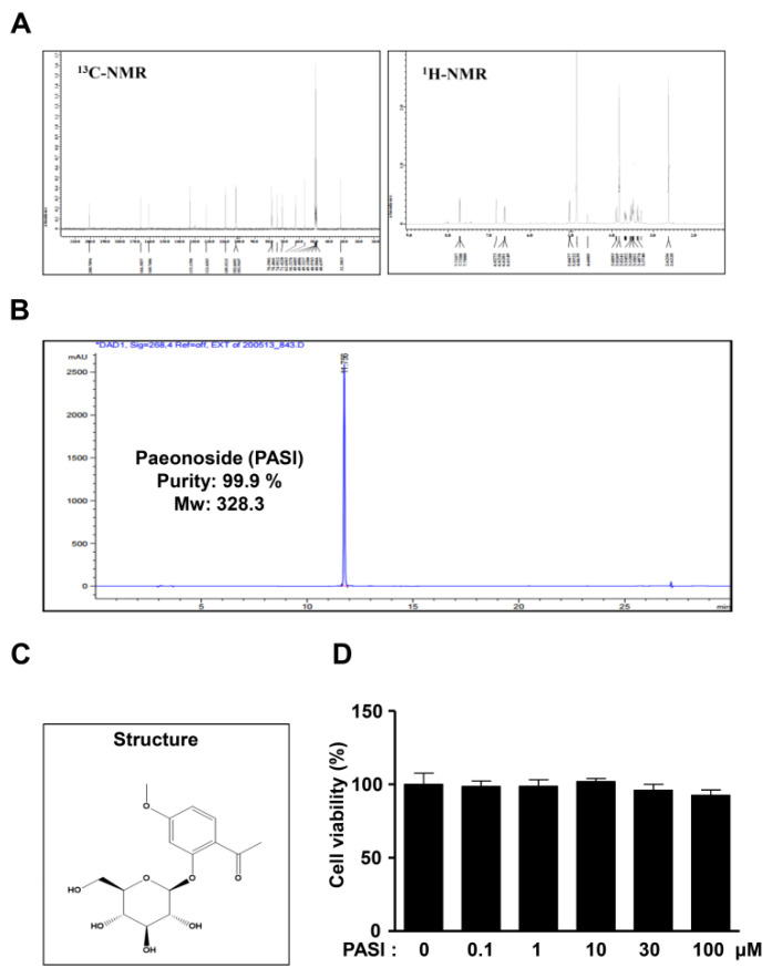
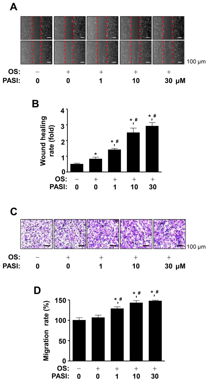
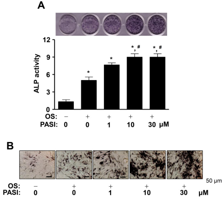
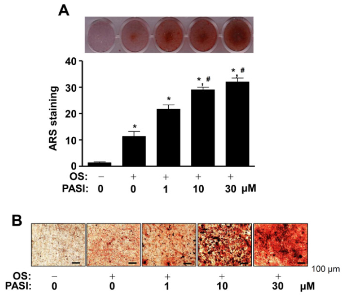
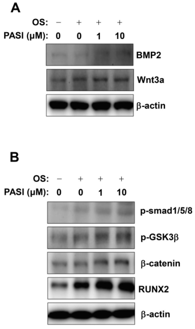
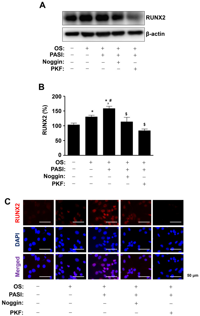
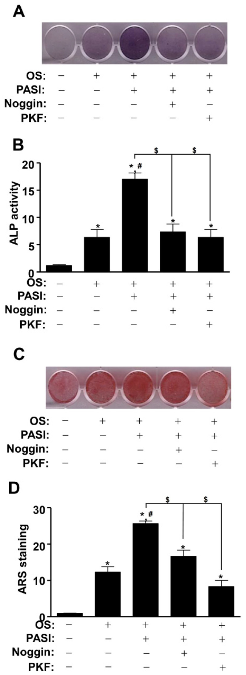
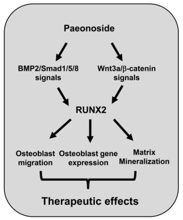
Paeonia suffruticosa is a magnificent and long-lived woody plant that has traditionally been used to treat various diseases including inflammatory, neurological, cancer, and cardiovascular diseases. In the present study, we demonstrated the biological mechanisms of paeonoside (PASI) isolated from the dried roots of P. suffruticosa in pre-osteoblasts. Herein, we found that PASI has no cytotoxic effects on pre-osteoblasts. Migration assay showed that PASI promoted wound healing and transmigration in osteoblast differentiation. PASI increased early osteoblast differentiation and mineralized nodule formation. In addition, PASI enhanced the expression of Wnt3a and bone morphogenetic protein 2 (BMP2) and activated their downstream molecules, Smad1/5/8 and β-catenin, leading to increases in runt-related transcription factor 2 (RUNX2) expression during osteoblast differentiation. Furthermore, PASI-mediated osteoblast differentiation was attenuated by inhibiting the BMP2 and Wnt3a pathways, which was accompanied by reduction in the expression of RUNX2 in the nucleus. Taken together, our findings provide evidence that PASI enhances osteoblast differentiation and mineralized nodules by regulating RUNX2 expression through the BMP2 and Wnt3a pathways, suggesting a potential role for PASI targeting osteoblasts to treat bone diseases including osteoporosis and periodontitis.

Keywords: BMP2; RUNX2; Wnt3a; bone mineralization; osteoblast differentiation; paeonoside.


Conflict of interest statement

The authors declare no conflict of interest.

Figures

Figure 1
Figure 1
Effects of PASI on cytotoxicity against pre-osteoblasts. (AC) PASI (99.9 % purity) obtained from the dried roots of Paeonia suffruticosa was analyzed by 1H and 13C NMR spectra (A), HPLC chromatogram (B), and chemical structure (C). (D) Cells were treated with 0.1–100 μM PASI for 24 h. Cell viability (%) was measured by using an MTT assay. Data are expressed as the mean ± S.E.M. of experiments.
Figure 2
Figure 2
Effects of PASI on migration during osteoblast differentiation. (A,B) The wound healing rate (fold) was detected under a light microscope (A) and exhibited as a bar graph (B). (C,D) The Boyden chamber assay was carried out. The migration rate (%) was detected under a light microscope (C) and exhibited as a bar graph (D). Data are expressed as the mean ± S.E.M. of experiments. * p < 0.05, # p < 0.05 indicate a statistically significant difference compared to the control and OS, respectively.
Figure 3
Figure 3
Effects of PASI on the staining and activity of ALP during early osteoblast differentiation. (A) The staining of ALP was visualized using a scanner (upper). The activity of ALP was analyzed by using a spectrophotometer and exhibited as a bar graph (bottom). (B) ALP-positive cells were detected under light microscopy. Data are expressed as the mean ± S.E.M. of experiments. * p < 0.05, # p < 0.05 indicate a statistically significant difference compared to the control and OS, respectively.
Figure 4
Figure 4
Effects of PASI on the staining of ARS during late osteoblast differentiation. (A) The staining of ARS was visualized using a scanner (upper). The quantification of ARS stains was analyzed by using a spectrophotometer and exhibited as a bar graph (bottom). (B) The formation of the nodules was detected under a light microscope. Data are expressed as the mean ± S.E.M. of experiments. * p < 0.05, # p < 0.05 indicate a statistically significant difference compared to the control and OS, respectively.
Figure 5
Figure 5
Effects of PASI on BMP2 and Wnt3a/β-catenin signaling during osteoblast differentiation. (A,B) The expression of BMP2 and Wnt3a proteins (A), and the phosphorylation of Smad1/5/8 (p-Smad1/5/8) and GSK3β (p-GSK3β) proteins, and the expression of β-catenin protein (B) were analyzed using western blot analysis. β-actin was used as an internal control to normalize the level of total lysates.
Figure 6
Figure 6
PASI promotes the expression of nuclear RUNX2 via BMP2 and Wnt3a/β-catenin signaling during osteoblast differentiation. (A,B) The expression of RUNX2 protein was analyzed using western blot analysis. β-actin was used as an internal control to normalize the level of total lysates (A). The expression levels of RUNX2 (%) are presented as a bar graph (B). (C) The nuclear expression of RUNX2 was examined using immunefluorescence analysis. The nucleus was stained with a nuclear DAPI marker. RUNX2-and DAPI-positively-stained cells were merged (purple, bottom). Data are expressed as the mean ± S.E.M. of experiments. * p < 0.05, # p < 0.05, and $ p < 0.05 indicate a statistically significant difference compared to the control, OS, and OS + PASI, respectively.
Figure 7
Figure 7
Blockage of BMP2 and Wnt3a/β-catenin signaling attenuated PASI-mediated osteoblast differentiation. (A,B) The staining of ALP was visualized using a scanner (A), and the activity of ALP was analyzed using a spectrophotometer and exhibited as a bar graph (B). (C,D) The staining of ARS was visualized using a scanner (C), and the quantification was presented as a bar graph (D) (bottom). Data are expressed as the mean ± S.E.M. of experiments. * p < 0.05, # p < 0.05, and $ p < 0.05 indicate a statistically significant difference compared to the control, OS, and OS + PASI, respectively.
Figure 8
Figure 8
Schematic showing PASI-mediated osteoblast differentiation and mineralization.

References

    1. Feng X., McDonald J.M. Disorders of bone remodeling. Annu. Rev. Pathol. 2011;6:121–145. doi: 10.1146/annurev-pathol-011110-130203. - DOI - PMC - PubMed
    1. Rucci N. Molecular biology of bone remodelling. Clin. Cases Miner. Bone Metab. 2008;5:49–56. - PMC - PubMed
    1. Marie P.J. Osteoblast dysfunctions in bone diseases: From cellular and molecular mechanisms to therapeutic strategies. Cell Mol. Life Sci. 2015;72:1347–1361. doi: 10.1007/s00018-014-1801-2. - DOI - PMC - PubMed
    1. Esfahanian V., Shamami M.S. Relationship between osteoporosis and periodontal disease: Review of the literature. J. Dent. 2012;9:256–264. - PMC - PubMed
    1. Fakhry M., Hamade E., Badran B., Buchet R., Magne D. Molecular mechanisms of mesenchymal stem cell differentiation towards osteoblasts. World J. Stem Cells. 2013;5:136–148. doi: 10.4252/wjsc.v5.i4.136. - DOI - PMC - PubMed

MeSH terms

LinkOut - more resources