Review
. 2021 Jun 27;13(13):3210.
doi: 10.3390/cancers13133210.

Epigenetic Deregulation of Apoptosis in Cancers

Affiliations
Review

Epigenetic Deregulation of Apoptosis in Cancers

Ezgi Ozyerli-Goknar et al. Cancers (Basel). .

Abstract

Cancer cells possess the ability to evade apoptosis. Genetic alterations through mutations in key genes of the apoptotic signaling pathway represent a major adaptive mechanism of apoptosis evasion. In parallel, epigenetic changes via aberrant modifications of DNA and histones to regulate the expression of pro- and antiapoptotic signal mediators represent a major complementary mechanism in apoptosis regulation and therapy response. Most epigenetic changes are governed by the activity of chromatin modifying enzymes that add, remove, or recognize different marks on histones and DNA. Here, we discuss how apoptosis signaling components are deregulated at epigenetic levels, particularly focusing on the roles of chromatin-modifying enzymes in this process. We also review the advances in cancer therapies with epigenetic drugs such as DNMT, HMT, HDAC, and BET inhibitors, as well as their effects on apoptosis modulation in cancer cells. Rewiring the epigenome by drug interventions can provide therapeutic advantage for various cancers by reverting therapy resistance and leading cancer cells to undergo apoptotic cell death.

Keywords: apoptosis; cancer; chromatin modifying enzymes; epigenetic drugs; evasion; therapy.


Conflict of interest statement

The authors declare no conflict of interest.

Figures

Figure 1
Figure 1
Morphological changes during apoptosis. Cells undergoing apoptosis start to shrink, which is followed by chromosome condensation and disintegration of organelles. Cells later collapse into apoptotic bodies that are further eliminated by the immune system. Figure generated at Biorender.com accessed on 20 April 2021.
Figure 2
Figure 2
Extrinsic and intrinsic apoptosis and its deregulation in cancer cells. (a) Extrinsic apoptosis inducers, such as TRAIL or FasL, bind to death receptors and lead to FADD-mediated caspase-8 activation. Active caspase-8 cleaves and activates effector caspase-3/7 and leads to apoptosis. Caspase-8 also truncates Bid and causes Bax and Bak oligomerization in mitochondrial outer membrane that leads to cytochrome C release, consequent activation of caspase-9 and effector caspases-3/7, and apoptosis (intrinsic). Intrinsic apoptosis can also be triggered by BH3-only proteins, which inhibit Bcl-2 and Bcl-xL antiapoptotic proteins, facilitating Bax and Bak activity. XIAP and cFLIP inhibit apoptosis through interfering with caspase activation. (b) Mechanisms of apoptosis evasion in cancer cells. The balance of pro- and antiapoptotic signal mediators is deregulated both transcriptionally (e.g., DNA hyper/hypomethylation) and post-translationally (e.g., phosphorylation) in cancer cells. Figure generated at Biorender.com accessed on 20 April 2021.
Figure 3
Figure 3
Subgroups of chromatin modifier proteins: writers, erasers, and readers. Post-translational marks are added by “writers”, recognized and further processed by “readers”, and removed by “erasers”. Figure generated at Biorender.com accessed on 20 April 2021.
Figure 4
Figure 4
Epigenetic modulation of gene expression through chromatin remodeling. Octamer sliding and DNA looping change the accessible surface area of nucleosomal DNA and regulate the access of transcription factors. Histone variants have distinct functions that could activate or repress gene expression in a context-dependent manner. Figure generated at Biorender.com accessed on 20 April 2021.
Figure 5
Figure 5
CpG island methylation blocks transcription. Methylation blocks the access of transcription factors to CpG sites and, therefore, results in transcriptional silencing. Figure generated at Biorender.com accessed on 20 April 2021.
Figure 6
Figure 6
Evasion of apoptosis by DNA hypermethylation and global hypomethylation. CpG islands at promoters of tumor suppressor, proapoptotic, cell-cycle regulator, and DNA damage repair genes are mostly hypermethylated in cancer cells due to DNMT overexpression or gene mutations, which lead to uncontrolled division and growth of cells. Cancer-associated hypomethylation occurs in several tumor-initiator or proliferation-associated genes and leads to chromosome instability. Figure generated at Biorender.com accessed on 20 April 2021.
Figure 7
Figure 7
Aberrant histone modifications and their contribution to evasion of apoptosis. Histone marks related to Table 3. and H4 due to high HDAC or low HAT activity, decreased H3K4me3 mark, and increased H3K9me3 and H3K27me3 modifications, which can silence tumor suppressor genes or proapoptotic genes to facilitate uncontrolled proliferation of cells. Histone phosphorylation and incorporation of histone variants also contribute to tumorigenesis by promoting proliferation and genomic instability. Figure generated at Biorender.com accessed on 20 April 2021.
Figure 8
Figure 8
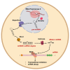
miRNA generation and action in the cell. miRNAs are small noncoding RNAs endogenously expressed in the cell and generated through a multistep process. RNA polymerase II synthe Table 5. pre-miRNAs are transported to the cytoplasm and get cleaved in the cytoplasm by Dicer into mature miRNA duplexes. Upon separation of the duplex, the guide strand gets loaded into the RISC complex and scans the transcriptome for complementary sites. Upon binding to complementary mRNA, RISC initiates either mRNA degradation or translation repression to control gene expression. Figure generated at Biorender.com accessed on 20 April 2021.

References

    1. Green D.R. Means to an End: Apoptosis and Other Cell Death Mechanisms. Cold Spring Harbor Laboratory Press; Cold Spring Harbor, NY, USA: 2010.
    1. Wyllie A.H., Kerr J.F.R., Currie A.R. Cell Death: The Significance of Apoptosis. Int. Rev. Cytol. 1980;68:251–306. doi: 10.1016/S0074-7696(08)62312-8. - DOI - PubMed
    1. Hanahan D., Weinberg R.A. The Hallmarks of Cancer Review Douglas. Cell. 2000;100:57–70. doi: 10.1016/S0092-8674(00)81683-9. - DOI - PubMed
    1. Fernald K., Kurokawa M. Evading apoptosis in cancer. Trends Cell Biol. 2013 doi: 10.1016/j.tcb.2013.07.006. - DOI - PMC - PubMed
    1. Esteller M. Epigenetics in cancer. N. Engl. J. Med. 2008;358:1148–1159. doi: 10.1056/NEJMra072067. - DOI - PubMed

LinkOut - more resources