Case Reports
. 2021 Jun 27;57(7):660.
doi: 10.3390/medicina57070660.

Cardiac Amyloidosis with Discordant QRS Voltage between Frontal and Precordial Leads

Affiliations
Case Reports

Cardiac Amyloidosis with Discordant QRS Voltage between Frontal and Precordial Leads

Csilla-Andrea Eötvös et al. Medicina (Kaunas). .

Abstract

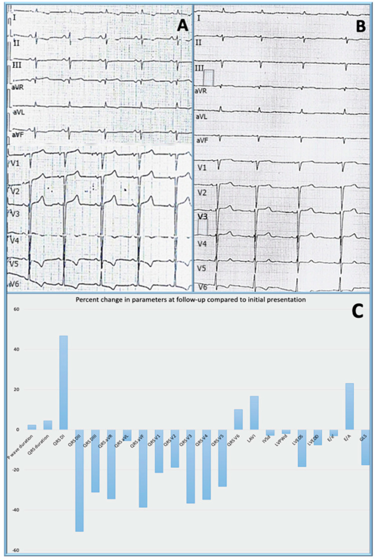
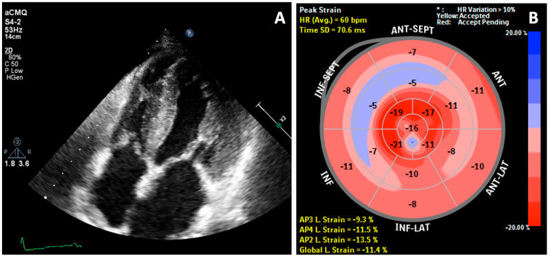
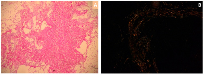
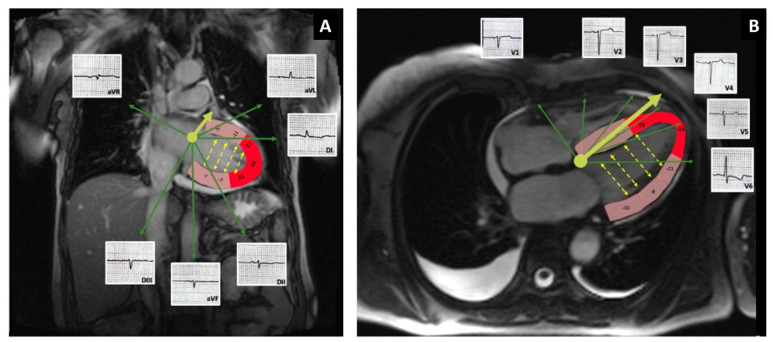
Among the different types, immunoglobulin light chain (AL) cardiac amyloidosis is associated with the highest morbidity and mortality. The outcome, however, is significantly better when an early diagnosis is made and treatment initiated promptly. We present a case of cardiac amyloidosis with left ventricular hypertrophy criteria on the electrocardiogram. After 9 months of follow-up, the patient developed low voltage in the limb leads, while still maintaining the Cornell criteria for left ventricular hypertrophy as well. The relative apical sparing by the disease process, as well as decreased cancellation of the opposing left ventricular walls could be responsible for this phenomenon. The discordance between the voltage in the frontal leads and precordial leads, when present in conjunction with other findings, may be helpful in raising the clinical suspicion of cardiac amyloidosis.

Keywords: amyloidosis; cardiomyopathy; low voltage.


Conflict of interest statement

The authors declare no conflict of interest.

Figures

Figure 1
Figure 1
Electrocardiogram at baseline (A) and at follow-up (B) and percent changes in electrocardiographic and echocardiographic parameters at 9 months follow-up compared to the initial presentation; QRS DI − QRS V6 = QRS voltage in all 12 leads of the standard electrocardiogram; LAVI = left atrial volume index; IVSd = interventricular septum in diastole; LVPWd = left ventricular posterior wall in diastole; LVEDS = left ventricular end-diastolic diameter in systole; LVEDD = left ventricular end-diastolic diameter in diastole; E/e′ = mitral E/e′ ratio; E/A = mitral ratio between the maximal velocity of transmitral flow in early to late diastole; GLS = global longitudinal strain, presented as change in absolute value (C); QRS voltage measured as average of three consecutive beats, using digital calipers at 300% magnification calibrated for paper speed of 25 mm/s.
Figure 2
Figure 2
2D Transthoracic echocardiography: (A) Thickening of both atria, right ventricle walls, atrial septum, valves, and papillary muscles and (B) Reduced global longitudinal strain (GLS = −11.4%) with bull’s eye plot and pathognomonic pattern of relative apical sparing.
Figure 3
Figure 3
Abdominal fat pad biopsy: hematoxylin and eosin-stained biopsy sections revealing eosinophilic amorphous amyloid fibrils (A) and extracellular amyloid deposits with typical apple-green birefringence with Congo red dye under polarized light microscopy (B).
Figure 4
Figure 4
Potential mechanism responsible for the discordance in voltage between frontal (A) and precordial leads (B). Fused cMRI-GLS images with examples of ECG waves on leads V1–V6. Segments of the LV are color-coded based on local strain values: red corresponds to local strain ≤−16%; pink local strain >−16%. The solid yellow arrow represents the QRS axis. The dotted yellow arrows represent direction of cancellation of electrical forces between opposing walls. The relative apical sparing pattern could be related to the high voltage seen in the precordial leads.

References

    1. Ablasser K., Verheyen N., Glantschnig T., Agnetti G., Rainer P.P. Unfolding cardiac amyloidosis—From pathophysiology to cure. Curr. Med. Chem. 2019;26:2865–2878. doi: 10.2174/0929867325666180104153338. - DOI - PubMed
    1. Martinez-Naharro A., Hawkins P.N., Fontana M. Cardiac amyloidosis. Clin. Med. (Lond.) 2018;18:s30–s35. doi: 10.7861/clinmedicine.18-2-s30. - DOI - PMC - PubMed
    1. Hirata Y., Kusunose K., Miki H., Yamada H. Improvement of global longitudinal strain following high-dose chemotherapy and autologous peripheral blood stem cell transplantation in patients with amyloid light-chain cardiac amyloidosis: A case report. Eur. Heart J. Case Rep. 2019;3:1–6. doi: 10.1093/ehjcr/ytz225. - DOI - PMC - PubMed
    1. Kristen A.V. Amyloid cardiomyopathy. Herz. 2020;45:267–271. doi: 10.1007/s00059-020-04904-4. - DOI - PubMed
    1. Maurea N., Riva L. Venous thromboembolism and atrial fibrillation in patients with cancer. G Ital. Cardiol. (Rome) 2018;19:3S–6S. doi: 10.1714/2989.29921. - DOI - PubMed

Publication types